
Contents
- Description
- Structure and function
- Epidemiology
- Clinical presentation
- Red flags
- Differential diagnosis
- Objective evidence
- Risk factors and prevention
- Treatment options
- Outcomes
- Miscellany
- Key terms
- Skills
Description
|
Scapula fractures are closely associated with high-energy trauma and often accompanied by numerous comorbid injuries, which are directly responsible for the patient prognosis. Consequently, fracture diagnosis is often delayed due to associated injuries. The most common scapular fractures are of the body, followed by the neck, glenoid, and acromion. |
Structure and function
|
The scapula is a flat, triangular bone that links the axial skeleton to the upper extremity via only muscular attachments. The scapula provieds the origin and mechanical leverage for the deltoid muscle, the prime mover of the shoulder. There are two palpable, bony landmarks, the acromion and the coracoid process.
The scapula has two true articulations. The glenohumoral joint is a highly mobile spheroidal ball and socket joint that requires the four muscles of the rotator cuff (supraspinatus, infraspinatous, teres minor, and subscapularis) for stability. This joint has a greater range of motion than any other joint. The acromioclavicular joint is a plane/gliding joint with a capsule and a disk in joint. The acromion has two ossification centers that fuse by age 22 years. Motion at this joint occurs in three planes: Anterior-Posterior gliding of acromion during protraction & retraction of scapula; tilting of acromion during abduction & adduction of arm; rotation of the clavicle during abduction & adduction of shoulder. The surrounding muscle mass (deltoid, trapezius, supraspinatus, and infraspinatus muscles) and the mobility of the scapula on the axial skeleton aide in force dissipation and provide protection from an impact injury. |


Epidemiology
|
Scapula fractures are relatively uncommon, accounting for only 3% to 5% of all shoulder fractures and 0.5% to 1% of all fractures. The average age of patients with scapula fractures is 35 to 45 years, but this injury is also associated with young males, due to its relationship to high-energy trauma (i.e. – motor vehicle accidents). |
Clinical presentation
|
Typically, significant high-energy trauma is required to fracture the scapula, with motor vehicle accidents accounting for approximately 50% of cases and motorcycle accidents in 11% to 25%. Trauma may occur directly from a hit or fall to the point of the shoulder (acromion, coracoid fracture) or indirectly through axial loading on the outstretched arm (scapular neck, glenoid, intra-articular fracture). The patient typically presents with the upper extremity supported by the contralateral hand in an immobile and adducted position with painful range of motion, especially with abduction. When a scapula fracture is suspected, the clinician should be suspicious of comorbid injuries and a full trauma evaluation is indicated, with focus on airway, breathing, circulation, disability, and exposure. After careful examination for associated injuries, a through neurovascular exam should follow. In the presence of pain out of proportion to the exam, compartment syndrome overlying the scapula, although uncommon, must be ruled out. A triangular swelling overlying the scapula, called Comolli sign, is suggestive of hematoma resulting in increased compartment pressures. |
Red flags
|
Diagnosis is often delayed due to comorbid injuries, thus suspicion for scapula fractures should arise when a patient presents with trauma and 1st rib fracture, pneumothorax, pulmonary contusion, pulmonary or great vessel injury, or acromioclavicular injury |
Differential diagnosis
|
Considering trauma is the primary mechanism of injury, any obvious pathology discovered in the trauma assessment should focus the clinician to structures that are associated with high-energy trauma. Fractured ribs (particularly 1st rib), clavicle, sternum, and proximal humerus trauma are often associated with ipsilateral scapula fractures. Breathing assessment may reveal pneumothorax and pulmonary contusion in 11% to 54% of scapula fractures. Hemodynamic instability with associated injuries to the pulmonary and great vessels or the brachiocephalic or subclavian may be seen. Brachial plexus injuries may be discovered on neurologic assessment, particularly suprascapular nerve deficits, such as loss of initial arm abduction via supraspinatus or loss of external rotation via infraspinatus. Comorbid spine injuries are common, with 76% thoracic spine, 20% lower cervical, and 4% lumbar spine. |
Objective evidence
|
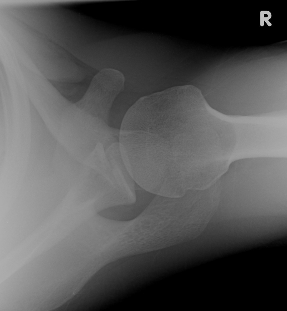
A scapula fracture may first be discovered during an essential part of any trauma evaluation: a chest x-ray. Suspicion of a scapula fracture should prompt a trauma series of the shoulder, consisting of a true anterioposterior (A/P) view, an axillary view, and a scapular-Y view (true scapular lateral). The axillary view is used to differentiate acromial and glenoid fractures. A 45-degree cephalic tilt (Stryker notch) radiograph is useful to identify coracoids fractures. Systematic review of the scapula includes the following:
|



Risk factors and prevention
|
Motor vehicle accidents and other high-energy trauma are responsible for up to 75% of scapula fractures. Proper use of safety belts and obeying traffic laws are paramount to avoiding these situations. |
Treatment options
|
Most extra-articular scapula fractures are responsive to non-operative treatment, such as sling use and early movement of shoulder through its range of motion. Scapular body fractures are generally responsive to non-operative treatment, unless when neurovascular compromise is present. Glenoid neck fractures can generally be treated with early range of motion exercises.
|
Outcomes
|
With early intervention and treatment, scapula fractures are amenable to a full recovery with restoration of the original range of motion and upper extremity strength prior to the injury. The associated injuries account for the majority of the morbidity and mortality. An increased mortality is associated with simultaneous 1st rib fracture. Fractures treated with non-operative treatment generally unite, but the risk of a malunion is present. This is generally well tolerated, but may result in scapulothoracic crepitus and localized pain |
Miscellany
A favorite “pimp” question on orthopedic rounds is the number of muscles that attach to the scapula.
The 18 muscles that attach to the scapula are summarized in Table 1.
|
Muscle |
Direction |
Region |
|
Pectoralis Minor |
insertion |
|
|
Coracobrachialis |
origin |
|
|
insertion |
||
|
Triceps Brachii (long head) |
origin |
|
|
Biceps Brachii (short head) |
origin |
|
|
Biceps Brachii (long head) |
origin |
|
|
Subscapularis |
origin |
|
|
insertion |
||
|
insertion |
||
|
insertion |
||
|
Trapezius |
insertion |
|
|
origin |
||
|
origin |
supraspinous fossa |
|
|
Infraspinatus |
origin |
infraspinous fossa |
|
Teres Minor |
origin |
lateral border |
|
Teres Major |
origin |
lateral border |
|
Latissimus Dorsi (a few fibers) |
origin |
inferior angle |
|
Omohyoid |
origin |
Table 1. The muscular attachments of the scapula.
Key terms
|
Scapula; glenoid; high-energy trauma; 1st rib fracture; pneumothorax |
Skills
|
Look for scapula fractures on any chest x-ray from a trauma; build a differential diagnosis for trauma victims that incorporates damage to the great vessels, pneumothorax and other pulmonary injuries, and structural injuries; order a trauma series of the shoulder, including: true AP, axillary, and true lateral views on any trauma victim suspected of injury; understand the indications for operative treatment versus non-operative treatment |