
1. High-Yield Overview & Pathognomonic Essence
The cotton wool sign is the classical, highly specific radiographic manifestation of late active and mixed-phase Paget’s disease of bone (osteitis deformans) involving the cranial vault:
- Fundamental Radiographic Appearance: Marked, irregular thickening of the calvarium with expansive diploic widening, indistinct demarcation of inner and outer cortical tables, and multifocal, coalescing, ill-defined rounded or nodular radiodensities (“cotton balls” or “cotton wool”) scattered across underlying areas of osteopenia.
- Pathognomonic Pathology: Represents the hallmark of advanced cranial Paget’s disease during the transition from active osteolysis to chaotic osteoblastic bone formation, where disorganized woven bone is deposited in an uncoupled, mosaic architectural pattern within the vascularized diploe.
- Critical Diagnostic & Clinical Implication: Readily confirms cranial involvement in Paget’s disease, differentiating it from osteoblastic skeletal metastases and multiple myeloma, while immediately signaling the clinician to screen for progressive auditory deficits, cranial nerve compression, basilar invagination, and the need for antiresorptive pharmacotherapy.
2. Diagnostic Sign Summary Matrix
| Primary Modality | Anatomic Location | Primary Pathology | Pathognomonic Morphology | Diagnostic Utility & Sensitivity |
|---|---|---|---|---|
| Plain Radiography (Lateral & AP Skull) | Cranial Vault (Frontal, Parietal, Occipital Bones) | Paget’s Disease of Bone (Mixed to Quiescent Phase) | Calvarial thickening, diploic widening, loss of cortical table distinctness, and multiple fluffy, coalescent rounded radiodensities (“cotton wool spots”) | First-line screening modality; specificity >95% in established mixed cranial Paget’s disease; diagnostic gold standard |
| Computed Tomography (HRCT) | Calvarium, Skull Base, and Temporal Bones | Cranial Paget’s Remodeling & Neuroforaminal Encroachment | Trabecular coarsening, expansive diploic thickening with mixed osteolysis and sclerosis; delineates petrous demineralization and canal narrowing | Superior sensitivity (>98%) for structural skull base anatomy, otic capsule involvement, foramen magnum encroachment, and microfractures |
| Magnetic Resonance Imaging (MRI) | Cranium, Diploic Space, and Craniocervical Junction | Diploic Bone Marrow Alterations & Neural Compression | Calvarial expansion with characteristic preservation of internal yellow/fatty marrow signal on T1; rules out soft tissue mass or cortical breakthrough | High specificity for ruling out osteosarcomatous transformation and osteoblastic metastasis; critical for evaluating basilar invagination and hindbrain herniation |
| Radionuclide Bone Scintigraphy (Tc-99m MDP) | Axial and Appendicular Skeleton (Whole Body) | Metabolically Active Osteoblastic Bone Turnover | Intense, asymmetric, diffuse or regionally expansive radiotracer uptake (“hot skull”) throughout the calvarium and base | Extreme sensitivity (>95-97%) for identifying active pagetic skeletal foci; crucial for establishing monostotic vs. polyostotic disease extent |
3. Clinical Case Imaging & Standardized Figures
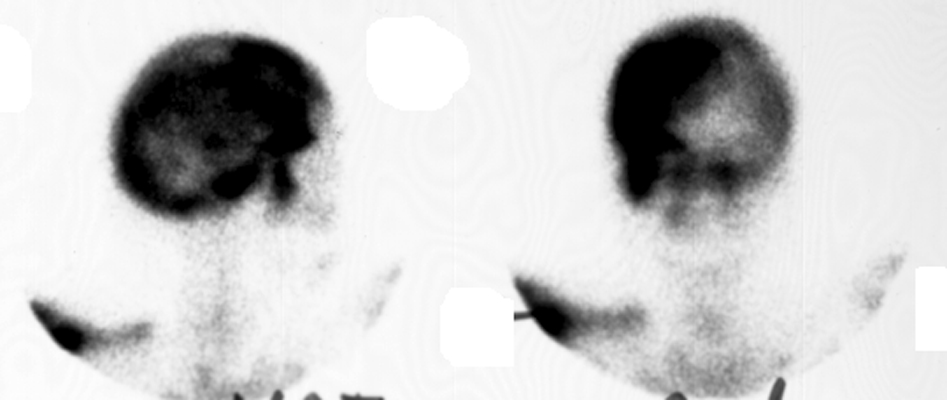
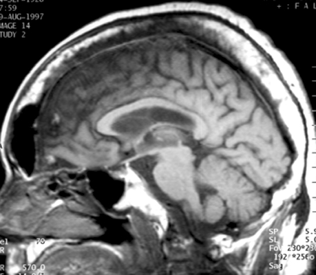
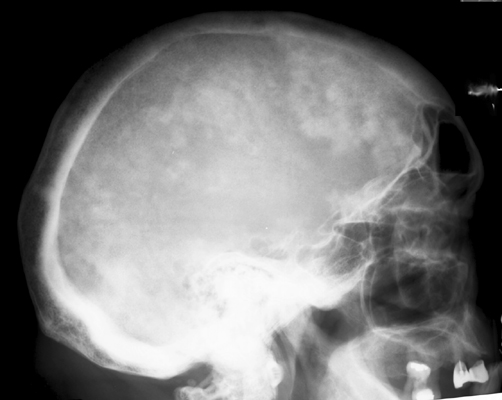
Comprehensive multimodality evaluation illustrated in Figures 1 through 3 encapsulates the complete morphological and functional spectrum of cranial Paget’s disease. The lateral plain radiograph (Figure 3) demonstrates the quintessential features of the cotton wool sign. The cranial vault exhibits marked calvarial expansion, with the diploic space widened to more than twice its physiologic thickness. The normally distinct inner and outer cortical tables are effaced and replaced by a chaotic architecture containing multiple irregular, cloud-like, fluffy radiodense islands that coalesce across a background of osteopenia. This appearance contrasts sharply with the sharply marginated radiolucency of the early osteolytic phase (osteoporosis circumscripta).
Cross-sectional sagittal T1-weighted magnetic resonance imaging (Figure 2) confirms pronounced osseous calvarial hypertrophy with dramatic expansion of the diploe. Crucially, the internal marrow space maintains predominantly high-to-intermediate signal intensity characteristic of preserved fatty marrow, interspersed with fibrous and sclerotic trabeculae. The complete absence of cortical erosion, soft-tissue mass extension, or dural invasion provides definitive evidence against secondary osteosarcomatous transformation or metastatic disease. Complementing these structural studies, Tc-99m MDP bone scintigraphy (Figure 1) demonstrates avid, intense radiotracer uptake throughout the cranial vault, with marked regional asymmetry. This scintigraphic hyperactivity confirms ongoing, accelerated bone turnover and hyperemic osteoblastic activity within the affected calvarium.
4. Pathophysiology & Biomechanical Mechanism
The cotton wool sign represents the radiographic culmination of a chronic, localized bone remodeling disorder driven by dysregulated osteoclastic hyperactivity coupled with accelerated, disorganized osteoblastic osteogenesis. Normal bone homeostasis relies on tightly orchestrated coupling between bone resorption and lamellar bone formation within basic multicellular units (BMUs). In Paget’s disease of bone, this coupling breaks down completely, initiating a characteristic triphasic pathological cascade.
The primary cellular defect resides in the osteoclast lineage. Pagetic osteoclasts are markedly abnormal: they are gigantic, containing up to 100 nuclei (compared to 3 to 8 in physiological osteoclasts), hyper-responsive to receptor activator of nuclear factor kappa-B ligand (RANKL) and 1,25-dihydroxyvitamin D3, and resistant to apoptosis. Genetic investigations have identified germline mutations in the SQSTM1 gene, encoding the scaffold protein sequestosome 1 (p62), in a substantial subset of familial and sporadic cases. This mutation leads to constitutive hyperactivation of the NF-kB signaling cascade. Ultrastructural studies have also demonstrated paramyxovirus-like nucleocapsid nuclear inclusions within osteoclasts, suggesting a potential gene-environment interaction involving slow viral infection (such as measles or canine distemper virus).
The disease progresses through three distinct, overlapping evolutionary stages in the cranial vault:
- Incipient Osteolytic Phase (Osteoporosis Circumscripta): Pathological resorption begins in the outer cortical table and diploe, typically arising in the frontal or occipital calvarium. Massive waves of giant osteoclasts excavate extensive geographic areas of bone matrix. On radiographs, this presents as sharply circumscribed, rounded radiolucent areas termed osteoporosis circumscripta cranii. The advancing front mimics a “blade of grass” or “candle flame” cutting cone seen in pagetic long bones.
- Mixed Osteolytic-Osteoblastic Phase (Origin of the Cotton Wool Sign): As osteolytic resorption continues, a compensatory, frenetic osteoblastic response is recruited. However, osteoblastic matrix synthesis is accelerated and unregulated. The newly synthesized collagen matrix is deposited in a haphazard, woven arrangement rather than organized lamellar sheets. Interspersed islands of woven bone undergo variable, incomplete mineralization within a hypervascular, cellular fibrovascular stroma. On plain radiography and CT, these nodular, disorganized foci of newly formed woven bone produce the fluffy, coalescing radiodense opacities responsible for the classic cotton wool appearance.
- Quiescent Sclerotic Phase: Osteoclastic resorption gradually subsides while osteoblastic bone apposition slows, leaving an expanded, thickened, and densely sclerotic calvarial vault. Histologically, bone produced during this phase displays a pathognomonic “mosaic” or “jigsaw puzzle” pattern of irregular, scalloped cement lines (tessellated appearance) visible under polarized light microscopy, reflecting repeated, disorganized cycles of resorption and deposition without biomechanical orientation.
From a biomechanical standpoint, the enlarged pagetic calvarium represents a profound architectural paradox: although the skull vault is dramatically thickened and sclerotic, the bone is mechanically weak, soft, and brittle. Immature woven bone lacks the torsional and compressive strength of normal lamellar cortical bone, and the expanded diploe exhibits a reduced Young’s modulus of elasticity. Under the persistent mechanical load of the cranium and the upward axial resistance of the cervical spine, the softened skull base and vault undergo progressive plastic deformation. This leads to flattening of the skull base (platybasia) and superior displacement of the cervical spine into the skull base (basilar invagination), frequently causing foramen magnum stenosis and cranial nerve entrapment.
Additionally, pagetic bone marrow is intensely hypervascular. Extensive arteriovenous micro-shunts form within the fibrovascular marrow spaces, accompanied by marked dilatation of meningeal and scalp vessels. This hyperdynamic circulation generates local warmth over the calvarium (“hot skull”) and, in patients with extensive polyostotic skeletal involvement (greater than 15% to 35% of the skeleton), can provoke elevated cardiac output states.
5. Modality-Specific Imaging Findings
Plain Radiography
Plain radiography of the skull represents the historical gold standard and primary diagnostic screening tool. A complete cranial series includes lateral, anteroposterior (AP Caldwell), and Towne views:
- Calvarial Diploic Expansion: The calvarium demonstrates progressive widening of the diploic space, often expanding from its normal 4 to 7 mm thickness to 20 mm or more, resulting in marked enlargement of the cranial circumference.
- Loss of Cortical Differentiation: The distinct anatomical boundaries between the inner cortical table, central diploic space, and outer cortical table are completely obliterated, replaced by a homogeneous or mottled expansile bone texture.
- Fluffy Sclerotic Foci (Cotton Wool Appearance): Multiple, poorly defined, nodular radiodensities measuring 5 to 20 mm in diameter coalesce within the osteopenic, expanded diploe. These fluffy cotton-wool opacities typically predominate in the frontal and parietal vaults.
- Osteoporosis Circumscripta: Large, geographic, sharply demarcated radiolucent zones of osteolysis may coexist with or precede sclerotic cotton-wool nodules, most frequently involving the frontal and occipital squama.
- Structural Vault & Base Deformity: In advanced disease, plastic softening yields the “Tam O’Shanter” skull deformity, where the expanded, softened calvarium appears to sag over the skull base and facial bones. Basilar invagination is identified when the odontoid tip extends greater than 5 mm above Chamberlain’s line (palato-occipital line) or McGregor’s line.
Computed Tomography
High-resolution computed tomography (HRCT) with bone algorithm reconstructions provides exquisite anatomical depiction of complex cranial and skull base alterations:
- Trabecular Coarsening & Architectural Remodeling: Cross-sectional bone windows (Hounsfield units +400 to +1200) clearly delineate the transition from early cortical thinning to chaotic trabecular thickening, demonstrating the coarse, porous matrix characteristic of pagetic woven bone.
- Temporal Bone & Otic Capsule Evaluation: HRCT of the temporal bone is the gold standard for investigating pagetic hearing loss. It demonstrates demineralization of the dense labyrinthine bone of the cochlear capsule, micro-osteolytic remodeling of the petrous apex, and narrowing of the internal auditory canal (IAC) or stylomastoid foramen.
- Skull Base Foramina & Craniocervical Geometry: Multiplanar coronal and sagittal reconstructions accurately quantify luminal dimensions of the foramen magnum, jugular foramen, hypoglossal canal, and optic canals, confirming or excluding bony neural entrapment.
- Exclusion of Cortical Breach: CT confirms the intact, albeit expanded and porous, cortical shell, reliably excluding aggressive bone destruction, focal osteolysis, or soft-tissue mass formation that heralds malignant sarcomatous degeneration.
Magnetic Resonance Imaging & Ultrasound
Magnetic resonance imaging (MRI) is essential for interrogating marrow composition, assessing cranial nerve pathology, and evaluating the brain parenchyma and craniocervical junction:
- T1-Weighted Sequences (Marrow Fat Preservation): The pathognomonic MRI feature of uncomplicated cranial Paget’s disease is the preservation of yellow (fatty) bone marrow within the expanded diploe. High-to-intermediate T1 signal remains visible, interspersed with low-signal fibrous tissue and sclerotic trabeculae. Total replacement of normal marrow fat by homogeneous low T1 signal is abnormal and points strongly toward metastatic carcinoma, myeloma, or secondary sarcoma.
- T2-Weighted and STIR Sequences: Signal intensity on T2 and fat-suppressed sequences is characteristically heterogeneous. Active fibrovascular areas show hyperintense signal, whereas dense cortical bone and sclerotic islands produce signal voids.
- Post-Contrast T1 (Gadolinium): Active pagetic phases exhibit variable, patchy contrast enhancement reflecting hypervascular fibrovascular tissue. Diffuse, nodular, or mass-like extraosseous enhancement extending into the scalp or epidural space is strictly abnormal and suggests secondary sarcomatous degeneration.
- Craniocervical Junction & Hindbrain Evaluation: Sagittal T1/T2 imaging directly visualizes the odontoid tip relative to the brainstem and cervicomedullary junction, documenting cord compression, syringomyelia, or cerebellar tonsillar herniation (acquired Chiari malformation) secondary to basilar invagination.
- Ultrasonic Evaluation: While conventional ultrasound cannot penetrate the intact calvarial vault, high-resolution color and power Doppler sonography of the scalp and superficial temporal arteries is clinically useful in older patients presenting with new-onset temporal headaches to distinguish pagetic calvarial hypervascularity from giant cell (temporal) arteritis.
6. Diagnostic Utility, Sensitivity & Specificity
The diagnostic utility of the cotton wool sign is exceptionally robust in established cranial Paget’s disease. Published radiological validation studies have established key diagnostic accuracy metrics across modalities:
- Diagnostic Specificity: The classic cotton wool sign on plain skull radiography carries a specificity exceeding 95% to 98% for Paget’s disease of the cranial vault. When accompanied by calvarial expansion and diploic widening, it is virtually pathognomonic.
- Diagnostic Sensitivity: Plain radiography sensitivity varies according to the evolutionary phase: sensitivity is relatively modest (60% to 70%) during early isolated osteolytic stages (osteoporosis circumscripta), but increases to greater than 90% once mixed osteoblastic remodeling and sclerotic cotton-wool nodules coalesce.
- Scintigraphic Sensitivity: Technetium-99m MDP bone scintigraphy exhibits the highest overall sensitivity (95% to 98%) for detecting active skeletal foci in Paget’s disease. Increased tracer uptake frequently precedes radiographic cotton-wool changes by several months or years. However, scintigraphy has lower specificity (60% to 70%) and cannot differentiate Paget’s disease from osteoblastic metastases or fibrous dysplasia without correlation with plain radiographs or CT.
- Predictive Value of MRI Marrow Signal: Preservation of internal fatty marrow signal on T1-weighted MRI boasts a negative predictive value (NPV) exceeding 99% for excluding secondary osteosarcomatous transformation. Conversely, focal cortical destruction with a soft-tissue mass has a positive predictive value approaching 100% for secondary malignancy.
- Biochemical Correlation: Total serum alkaline phosphatase (ALP) and bone-specific alkaline phosphatase (BSAP) correlate directly with the volumetric burden of metabolically active pagetic bone. In patients with monostotic cranial Paget’s disease, marked elevations of ALP (often 2 to 5 times the upper limit of normal) are typical because of the substantial mass and hypervascularity of the cranial vault. Serum procollagen type I N-terminal propeptide (P1NP) and C-terminal telopeptide (beta-CTX) serve as sensitive secondary markers for monitoring therapeutic response to bisphosphonate therapy.
7. Differential Diagnosis & Radiographic Mimics
Differentiating the cotton wool sign from benign and malignant cranial lesions is essential for appropriate patient management and to prevent unnecessary invasive biopsies:
| Sign / Condition | Distinguishing Radiographic Features | Clinical Presentation | Key Differentiating Test |
|---|---|---|---|
| Cranial Paget’s Disease (Cotton Wool Sign) | Profound calvarial thickening, marked diploic expansion, effaced cortical tables, fluffy coalescent sclerotic islands; preserved T1 fatty marrow | Patients >55 years; progressive enlargement of the head (“hat sign”), sensorineural/conductive hearing loss, warm scalp, cranial bone pain | Lateral skull radiograph; elevated serum ALP; Tc-99m bone scan; preserved fatty marrow on T1-weighted MRI |
| Osteoblastic Bone Metastases (Prostate / Breast) | Multifocal, discrete, dense, rounded sclerotic lesions without calvarial expansion; sharp margins; normal diploic thickness; absence of osteoporosis circumscripta | Known primary adenocarcinoma; rapid onset of focal bone pain, weight loss, cachexia, constitutional symptoms | Complete replacement of T1 fatty marrow signal on MRI; elevated PSA or CA 15-3; cold/variable osteolysis; CT/PET imaging |
| Multiple Myeloma / Osteolytic Metastases | Multiple “punched-out,” sharply defined, non-sclerotic lytic lesions of variable size (“rain-drop skull”); no calvarial expansion; absence of sclerosis | Deep cranial pain, generalized fatigue, anemia, hypercalcemia, renal insufficiency (CRAB features), pathologic fractures | Serum and urine protein electrophoresis (SPEP/UPEP) with immunofixation; serum free light chains; bone marrow aspiration |
| Craniofacial Fibrous Dysplasia | Characteristic homogeneous “ground-glass” matrix expansion; thinning and ballooning of outer cortical table; predilection for anterior skull base, sphenoid, and facial bones | Children and young adults (<30 years); progressive painless facial asymmetry, proptosis, visual field defects; unilateral predominance | High-resolution CT displaying classic ground-glass matrix without fluffy cotton-wool nodules; GNAS1 gene mutation analysis |
| Hyperostosis Frontalis Interna | Symmetrical, lobulated, or nodular hyperostosis strictly confined to the inner table of the frontal bone; diploe and outer table completely preserved; sagittal sinus spared | Postmenopausal females; typically an asymptomatic, incidental radiographic finding; may be associated with Morgagni-Stewart-Morel syndrome | Skull radiography or CT demonstrating strictly inner table thickening with normal diploic thickness and normal alkaline phosphatase |
| Secondary Cranial Osteosarcoma (Malignant Transformation) | Focal zone of aggressive bone destruction within pagetic bone, cortical breakthrough, sunburst periosteal reaction, expanding soft-tissue mass | Known Paget patient presenting with sudden, intractable focal pain, rapid enlargement of a localized cranial mass, neurological decline | Contrast-enhanced MRI showing soft-tissue invasion and loss of fatty marrow; sudden exponential rise in serum ALP; urgent biopsy |
8. Clinical Significance & Management Implications
The radiographic identification of the cotton wool sign holds major clinical and therapeutic implications. Because the calvarium and skull base represent high-risk skeletal regions prone to irreversible neurological and structural complications, detection of this sign mandates a structured medical and surgical management strategy:
- Neurosensory Complications & Auditory Loss: Hearing loss is the most frequent clinical complication of cranial Paget’s disease, occurring in 30% to 50% of affected individuals. Hearing impairment may be conductive, sensorineural, or mixed. Sensorineural loss results from micro-architectural demineralization and loss of density within the otic capsule and cochlea, as well as microvascular steal from the internal auditory artery, rather than gross mechanical compression of the eighth cranial nerve. Baseline pure-tone audiometry and temporal bone HRCT are mandatory at the time of diagnosis.
- Cranial Neuropathies & Skull Base Compression: Progressive bony remodeling and narrowing of the skull base foramina can cause optic atrophy from optic canal stenosis, trigeminal neuralgia, facial weakness, and lower cranial nerve palsies (glossopharyngeal, vagus, and accessory nerves at the jugular foramen).
- Craniocervical Instability & Basilar Invagination: Structural softening of the occiput and sphenoid leads to platybasia and basilar invagination. The odontoid process migrates superiorly into the posterior fossa, compressing the medulla oblongata, upper cervical spinal cord, and anterior spinal arteries. This can produce quadriparesis, gait ataxia, nystagmus, hydrocephalus due to cerebral aqueduct obstruction, and life-threatening central sleep apnea.
- Medical Pharmacotherapy (First-Line Antiresorptive Therapy): Clinical practice guidelines from the Endocrine Society and the Journal of Bone and Mineral Research strongly recommend medical treatment for active cranial Paget’s disease, even in asymptomatic patients, to halt progressive bone remodeling and prevent permanent neurological and otological deficits. Intravenous zoledronic acid (a single 5 mg infusion administered over 15 minutes) is the first-line treatment of choice, providing rapid, sustained biochemical remission with normalization of serum ALP in over 90% to 95% of patients for up to 5 to 10 years. Oral bisphosphonates (risedronate 30 mg daily for 2 months, or alendronate 40 mg daily for 6 months) represent second-line alternatives. Serum 25-hydroxyvitamin D and calcium levels must be optimized prior to infusion to prevent profound hypocalcemia.
- Surgical Indications & Operative Precautions: Neurosurgical intervention is reserved for severe basilar invagination with symptomatic brainstem or cervical cord compression, refractory hydrocephalus (requiring ventriculoperitoneal shunting), or severe cranial nerve entrapment. Craniectomy and suboccipital decompression with C1-C2 stabilization are indicated when progressive myelopathy occurs. Crucially, pagetic bone is intensely hypervascular: craniotomy carries a formidable risk of massive intraoperative hemorrhage from expanded diploic veins and dural shunts. Preoperative administration of zoledronic acid at least 2 to 3 months prior to elective surgery markedly reduces bone vascularity and intraoperative blood loss. Meticulous local hemostatic techniques (bone wax, bipolar cautery, topical thrombin, and intraoperative cell salvage) are vital.
- Surveillance for Secondary Malignant Transformation: Secondary sarcomatous degeneration (osteosarcoma, undifferentiated pleomorphic sarcoma, or chondrosarcoma) occurs in approximately 0.5% to 1% of patients with long-standing Paget’s disease. The calvarium is a well-recognized site. Any new onset of localized severe cranial pain, rapid focal enlargement of a skull mass, or sudden spike in serum alkaline phosphatase warrants immediate cross-sectional imaging with contrast-enhanced MRI and prompt open biopsy.
9. Master Class Pro-Tip
Master Class Pro-Tip: When confronted with the classic cotton wool sign on a lateral skull radiograph, two critical diagnostic instincts must be engaged immediately. First, do not mistake cranial Paget’s disease for osteoblastic metastases: immediately review sagittal T1-weighted MRI sequences to confirm the preservation of internal fatty marrow signal within the thickened calvarium, which reliably excludes diffuse metastatic adenocarcinoma or multiple myeloma. Second, never dismiss cranial Paget’s disease as a benign, quiescent finding. Skull involvement is a high-risk anatomic site that dictates prompt baseline audiometry, temporal bone CT, and first-line treatment with intravenous zoledronic acid (5 mg) to arrest petrous bone demineralization before permanent sensorineural deafness or basilar invagination occurs. Finally, if elective craniotomy or surgical decompression is contemplated, initiate bisphosphonate therapy at least 8 to 12 weeks preoperatively to suppress the profuse diploic arteriovenous shunts and prevent catastrophic intraoperative hemorrhage.
Master Class Radiological Pearl: Cranial Paget’s Disease
10. Evidence-Based Synthesis
| Author & Year | Study Design | Sample Size | Key Findings & Diagnostic Accuracy | Clinical Implication | Level of Evidence | DOI |
|---|---|---|---|---|---|---|
| Ralston et al. (2019) | Clinical Practice Guideline & Systematic Review | Systematic review of 124 studies | Plain radiography is the gold standard for diagnosing Paget’s disease; radionuclide bone scintigraphy defines overall skeletal extent. Intravenous zoledronic acid (5 mg) is the primary first-line therapeutic agent to achieve durable biochemical remission and prevent skeletal complications. | Radiographic identification of the cotton wool appearance accompanied by elevated bone turnover markers warrants antiresorptive therapy to forestall progressive cranial and auditory complications. | Level I | DOI |
| Singer et al. (2014) | Clinical Practice Guideline | Endocrine Society Expert Consensus Panel | Recommends treatment of active Paget’s disease involving high-risk skeletal regions, specifically the skull and skull base, even in asymptomatic patients, to prevent irreversible cranial neuropathies, hearing loss, and basilar invagination. | Cotton wool sign involving the calvarium signifies high-risk skeletal anatomy requiring active antiresorptive management over observation. | Level I | DOI |
| Lentle et al. (1976) | Prospective Diagnostic Cohort Study | N = 65 patients | Demonstrated that Tc-99m bone scintigraphy has exceptional diagnostic sensitivity (>95%) for identifying metabolically active pagetic skeletal foci, frequently preceding clear-cut radiographic manifestations. | Baseline whole-body scintigraphy should accompany skull radiographs to distinguish monostotic from polyostotic disease distribution. | Level II | DOI |
| Monsell et al. (1999) | Prospective Observational Cohort Study | N = 77 cranial Paget patients | Established that hearing impairment directly correlates with demineralization and micro-architectural remodeling of the cochlear capsule and petrous temporal bone rather than gross mechanical compression of the internal auditory canal. | High-resolution computed tomography of the temporal bone is indicated when skull vault cotton wool signs are accompanied by auditory symptoms. | Level II | DOI |
| Smith et al. (2002) | Radiologic-Pathologic Correlation & Retrospective Case Series | N = 265 cases (AFIP archives) | Delineated the histopathologic basis of the cotton wool appearance: initial geographic osteolysis (osteoporosis circumscripta) transitions into chaotic, mosaic woven bone formation with osteoblastic apposition, producing patchy radiodense patches embedded within an expanded diploe. | Provides definitive radiological-pathological criteria for differentiating benign Paget’s disease from osteoblastic metastases and detecting secondary osteosarcomatous transformation. | Level IV | DOI |
| Whitten & Saifuddin (2003) | Retrospective Multimodality MRI Review | N = 78 patients | Confirmed that preservation of internal fatty marrow signal on T1-weighted MRI sequences across expanded calvarial bone is pathognomonic for uncomplicated Paget’s disease, distinguishing it from diffuse osteoblastic metastasis or myeloma. | Presence of soft-tissue mass, cortical destruction, or complete replacement of fatty marrow on MRI signifies secondary sarcomatous degeneration. | Level IV | DOI |
| Theodorou et al. (2011) | Comprehensive Multimodality Review | Multicenter imaging cohort review | Systematically categorized cranial manifestations and structural deformities in Paget’s disease, emphasizing platybasia, basilar invagination, and foramen magnum encroachment resulting from calvarial remodeling and softening. | Cross-sectional imaging (CT and MRI) is required to evaluate craniocervical junction geometry in advanced skull Paget’s disease. | Level V | DOI |
| Paget (1877) | Seminal Case Series & Historical Monograph | N = 5 patients | Original landmark description of osteitis deformans, detailing insidious enlargement of the skull vault leading to increased hat size, calvarial softening, and accompanying long bone deformities. | Foundational historical reference defining the clinical phenotype and natural history of osteitis deformans. | Level V | DOI |
11. References
- Ralston SH, Corral-Gudino L, Cooper C, et al. Diagnosis and Management of Paget’s Disease of Bone in Adults: A Clinical Guideline. J Bone Miner Res. 2019;34(4):579-604. DOI [Level I: Clinical Practice Guideline & Systematic Review]
- Singer FR, Bone HG, Hosking DJ, et al. Paget’s disease of bone: an Endocrine Society clinical practice guideline. J Clin Endocrinol Metab. 2014;99(12):4408-4422. DOI [Level I: Clinical Practice Guideline]
- Lentle BC, Russell AS, Heslip JM, Percy JS. The scintigraphic findings in Paget’s disease of bone. Clin Radiol. 1976;27(1):129-135. DOI [Level II: Prospective Diagnostic Cohort Study]
- Monsell EM, Cody DD, Bone HG, et al. Hearing loss as a complication of Paget’s disease of bone. J Bone Miner Res. 1999;14(2):224-231. DOI [Level II: Prospective Observational Cohort Study]
- Smith SE, Murphey MD, Motamedi K, Mulligan ME, Resnik CS, Gannon FH. From the archives of the AFIP. Radiologic spectrum of Paget disease of bone and its complications with pathologic correlation. RadioGraphics. 2002;22(5):1191-1216. DOI [Level IV: Radiologic-Pathologic Correlation & Retrospective Case Series]
- Whitten CR, Saifuddin A. MRI of Paget’s disease of bone. Clin Radiol. 2003;58(10):763-781. DOI [Level IV: Retrospective Multimodality MRI Review]
- Theodorou DJ, Theodorou SJ, Kakitsubata Y. Imaging of Paget disease of bone and its musculoskeletal complications: review. AJR Am J Roentgenol. 2011;196(6 Suppl):S64-S75. DOI [Level V: Comprehensive Multimodality Review]
- Paget J. On a form of chronic inflammation of bones (osteitis deformans). Medico-Chirurgical Transactions. 1877;60:37-63. DOI [Level V: Seminal Case Series & Historical Monograph]
Last Updated on September 29, 2026
🔗 Knowledge Graph & Clinical Correlates
OrthopaedicsOne Living Semantic Network🛠️ Operative Techniques
- Total scapular resections with endoprosthetic reconstruction Musculoskeletal Tumor
🩺 Clinical Conditions & Indications
- Who should undergo biopsy of a bone lesion Patient selection for a biopsy is important: It is intended to confirm the radiologic diagnosis in patients with...
- Who should do the biopsy of a bone lesion Clinical studies undertaken by the Musculoskeletal Tumor Society have documented a high incidence of problems suffered by patients...
- Where is the lesion Original Clinical Contribution: Authored for the OrthopaedicsOne Musculoskeletal Oncology Specialty Section. Preserved, digitized, and updated to modern evidence-based...
- What is the lesion doing to the bone Bone tumors have a limited number of potential effects on the skeleton. The most common is to produce...
🔬 Anatomy, Pathology & Diagnostics
- What kind of matrix is being made Generalized Bone and Joint
- Validation of cDNA Microarray Analysis to Distinguish Tumor Type Ex Vivo Generalized Bone and Joint
- Urokinase Plasminogen Activator System – A Multifunction Role in Tumour Progression and Metastasis Pelvis and Acetabulum
- Tumor Classifications Hip Joint & Proximal Femur