
Contents
- Name of bone
- Location/Articulation
- Muscle and ligament attachments
- Surface anatomy
- Radiography
- Physical examination
- Embryology
- Anomalies
- Injuries/Disorders
Name of bone
Femur
Location/Articulation
The femur, or thigh bone, is the most proximal (closest to the body) bone of the human leg. It articulates at the hip with the acetabulum of the pelvis superiorly and at the knee with the tibia and patella inferiorly.
Muscle and ligament attachments
Surface anatomy
The femur is the longest and largest bone, and one of the strongest bones in the body, along with the temporal bone of the skull. The average adult human femur is 48 cm (19 inches) in length and 2.34 cm (0.92 inches) in diameter; it can support up to 30 times the weight of an adult.
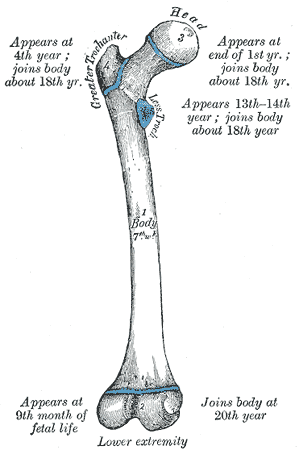
The femur forms part of the hip (at the acetabulum) and part of the knee. There are four eminences, or protuberances, in the human femur:
- head
- greater trochanter
- lesser trochanter
- lower extremity.
They appear at various times from just before birth to about age 14. Initially, these eminences are joined to the main body of the femur with cartilage, which gradually becomes ossified until the eminences become an integral part of the femur bone, usually in early adulthood.

Figure 1. Eminences of the femur
The intercondylar fossa is present between the condyles at the distal end of the femur. In addition to the intercondylar eminence on the tibial plateau, there is both an anterior and posterior intercondylar fossa (area), the sites of anterior cruciate and posterior cruciate ligament attachment, respectively.
Radiography
Physical examination
Embryology
Anomalies
Injuries/Disorders
Femur fracture.
Attachments:

Last Updated on June 27, 2024
🔗 Knowledge Graph & Clinical Correlates
OrthopaedicsOne Living Semantic Network🛠️ Operative Techniques
- Valgus osteotomy for Perthes disease Pediatrics
- Valgus osteotomy for development coxa vara Hip and Knee
🔬 Anatomy, Pathology & Diagnostics
- Wrist joint Wrist and Carpus
- Why I Would Not Consider Navigating My THA Hip Joint & Proximal Femur
- Why I Would Consider Navigating My THA Hip Joint & Proximal Femur
- Why I Use Metal on Metal Hip Joint & Proximal Femur