
Contents
- Name of bone
- Location/Articulation
- Muscle and ligament attachments
- Surface anatomy
- Ossification and Blood supply
- Radiography
- Physical examination
- Embryology
- Anomalies
- Injuries/Disorders
Name of bone
Fibula

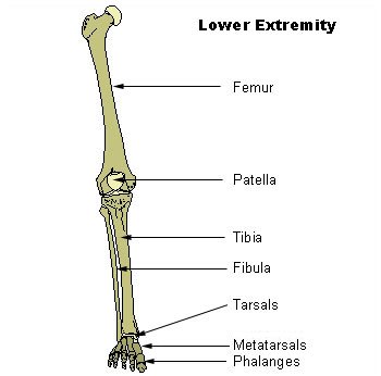
Figure 1. Bones of the lower extremity
Location/Articulation
The fibula, or calf bone, is located on the lateral side of the tibia, with which it is connected above and below. It is the smaller of the two bones, and, in proportion to its length, the most slender of all the long bones.
The upper extremity of the fibula is small, placed toward the back of the head of the tibia, below the level of the knee-joint, and excluded from the formation of this joint. Its lower extremity inclines a little forward, so as to be on a plane anterior to that of the upper end; it projects below the tibia, and forms the lateral part of the ankle-joint.
The term fibula derives from the Latin fibula, also meaning a clasp or brooch. The bone was so called because it resembles a clasp like a modern safety pin.
Muscle and ligament attachments
Surface anatomy
The fibula has the following components:
- Body
- Lateral malleolus
- Interosseous membrane connecting the fibula to the tibia, forming a syndesmoses joint
- The superior tibiofibular articulation is an arthrodial joint between the lateral condyle of the tibia and the – head of the fibula.
- The inferior tibiofibular articulation (tibiofibular syndesmosis) is formed by the rough, convex surface of the medial side of the lower end of the fibula, and a rough concave surface on the lateral side of the tibia.
Ossification and Blood supply
The fibula is ossified from three centers, one for the shaft and one for either end. Ossification begins in the body of the fibula about the 8th week of fetal life, and extends toward the extremities. At birth, the ends are cartilaginous. Ossification in the lower end of the fibula begins in the 2nd year; in the upper end, ossification begins about the 4th year. The lower epiphysis, the first to ossify, unites with the body about the 20th year; the upper epiphysis joins about the 25th year.
The blood supply of the fibula is important for planning free tissue transfer: the fibula is commonly used to reconstruct the mandible. The shaft is supplied in its middle third by a large nutrient vessel from the peroneal artery. It is also perfused from its periosteum, which receives many small branches from the peroneal artery. The proximal head and the epiphysis are supplied by a branch of the anterior tibial artery. In harvesting the bone, the middle third is always taken, with the ends preserved (4 cm proximally and 6 cm distally)
Radiography
Physical examination
Embryology
Anomalies
Injuries/Disorders
Attachments:

Last Updated on June 27, 2024
🔗 Knowledge Graph & Clinical Correlates
OrthopaedicsOne Living Semantic Network🛠️ Operative Techniques
- Upper Tibial Osteotomy Musculoskeletal Medicine
🔬 Anatomy, Pathology & Diagnostics
- Wrist joint Wrist and Carpus
- Vertebra prominens Surface Anatomy Lumbar Spine
- Vastus medialis Femoral Shaft
- Vastus lateralis Femoral Shaft