• O1 Network
    • Articles
    • For Residents
    • For Medical Students
    • PORT Notes
    • MSK Medicine
    • Tumor Library
    • Cases
    • Talks
    • Images
    • Viewpoints
    • Question Bank
    • Devices
    • Residency
    • Fellowships
    • Associations
    • Companies
ORTHOPAEDICSONE ARTICLES
OrthopaedicsOne Articles
  • Articles
    • Specialty
      • Arthroscopy
      • Foot and Ankle
      • Hand and Wrist
      • Hip and Knee
      • Infection
      • Joint Reconstruction
      • Musculoskeletal Medicine
      • Pediatrics
      • Shoulder and Elbow
      • Spine
      • Sports Medicine
      • Trauma
      • Tumor
    • Regional
      • Anatomy
      • Basic Science
      • Classifications
      • Diagnostic Tests
      • Evidence-Based Orthopaedics
      • Operative Techniques
      • Pain Management
      • Physical Examination
      • Pharmacology
      • Radiology
      • Rehabilitation
      • Rheumatology
      • Surgical Approaches
    • Professional
      • Bioethics
      • Economics and Health Policy
      • Epidemiology and Biostatistics
      • History of Orthopaedics
      • Informatics
      • Medicolegal
      • Mobile and Wireless
      • Practice Management
  • Reviews
    • For Residents
    • For Medical Students
  • eBooks
    • PORT Notes
    • MSK Medicine
    • Tumor Library
  • Community
    • Cases
    • Talks
    • Images
    • Viewpoints
    • Question Bank
    • Devices
  • Pages
    • Residency
    • Fellowships
    • Associations
    • Companies
  • About O1
    • Overview
    • FAQs
    • Managing Editors
    • Editorial Board
    • Content Partners
    • Participating Groups
    • Advisory Board
    • Sponsoring Organizations
    • Contact Us
  • Click to open the search input field Click to open the search input field Search
  • Menu Menu

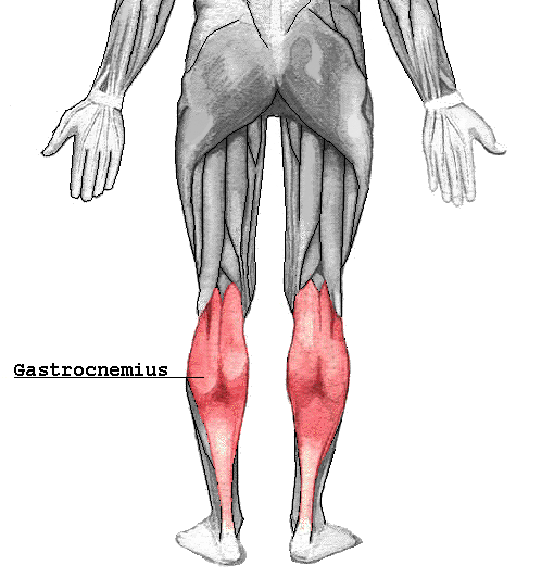
Gastrocnemius

Origin

Superior to articular surfaces of lateral condyle of femur and medial condyle of femur

Insertion

Tendo calcaneus (achilles tendon) into mid-posterior calcaneus

Action(s)

Plantar flexes foot, flexes knee

Nerve Supply

Tibial nerve from the sciatic, specifically, nerve roots S1–S2

Arterial Supply

Sural arteries

Physical Exam

Enter physical examination maneuvers for muscle

Clinical Importance

The gastrocnemius muscle is very prone to spasms; the painful, involuntary, contraction of the muscle for up to several minutes.

This muscle is prone to injury called torn calf muscle which is disabling.

The Gastrocnemius muscle may also become inflamed due to overuse. Anti-inflammatory and physical therapy may be necessary.

Anatomical abnormalities involving the medial head of gastrocnemius muscle results in popliteal entrapment syndrome.

Disease States

Enter links to pages where muscle involved

Discussion

The gastrocnemius is located with the soleus in the posterior (back) compartment of the leg. It originates from the posterior (back) surfaces of the distal head of the femur. Its other end forms a common tendon with the soleus muscle; this tendon is known as the calcaneal tendon or Achilles Tendon and inserts onto the posterior surface of the calcaneus, or mountain bone.

Deep to the gastrocnemius (farther from the skin) is the soleus muscle. Some anatomists consider both to be a single muscle, the triceps surae. The plantaris muscle and a portion of its tendon run between the two muscles, which is involved in “locking” the knee from the standing and posterior tibial vein and the tibial nerve. Since the anterior compartment of the leg is lateral to the tibia, the bulge of muscle medial to the tibia on the anterior side is actually the posterior compartment. The soleus is superficial midshaft of the tibia. Frequently there is a sesamoid bone called the “fabella” in the lateral head of gastrocnemius muscle.

Figures

Click thumbnail for larger image

Credits:

From Wikipedia:
Gastrocnemius

Attachments:


Gastrocnemius.png (image/png)

Last Updated on June 27, 2024

🔗 Knowledge Graph & Clinical Correlates

OrthopaedicsOne Living Semantic Network

🛠️ Operative Techniques

  • Use of Large Femoral Head Allograft for Tibiotalocalcaneal Arthrodesis Foot and Ankle
  • Triple Arthrodesis Foot and Ankle

🧭 Surgical Approaches & Danger Zones

  • U approach to the calcaneus Hindfoot (Talus & Calcaneus)

🔬 Anatomy, Pathology & Diagnostics

  • Wrist joint Wrist and Carpus
  • Vertebra prominens Surface Anatomy Lumbar Spine
  • Vastus medialis Femoral Shaft
  • Vastus lateralis Femoral Shaft

🕸️ Semantic Relations (RDF)

ABOUT

  • Overview
  • FAQs
  • Managing Editors
  • Editorial Board
  • Content Partners
  • Participating Groups
  • Advisory Board
  • Sponsoring Organizations
  • Contact Us

ARTICLES

  • Arthroscopy
  • Foot and Ankle
  • Hand and Wrist
  • Hip and Knee
  • Infection
  • Joint Reconstruction
  • Musculoskeletal Medicine
  • Pediatrics
  • Shoulder and Elbow
  • Spine
  • Sports Medicine
  • Trauma
  • Tumor

REVIEWS

  • For Residents
  • For Medical Students

EBOOKS

  • PORT Notes
  • MSK Medicine
  • Tumor Library

COMMUNITY

  • Talks
  • Images
  • Viewpoints
  • Question Bank
  • Devices

PAGES

  • Residency
  • Fellowships
  • Associations
  • Companies
Copyright © 2012-2026 OrthopaedicsOne | Privacy Policy | Dr. Christian Veillette . Powered by: OrthoNet - Empowering the Future of Orthopaedic Care
Scroll to top Scroll to top Scroll to top