
1. High-Yield Overview & Pathognomonic Essence
The Hill-Sachs sign represents a critical osseous lesion reflecting anterior glenohumeral dislocation and joint instability:
- Morphologic Appearance: A wedge-shaped compression fracture or grooved cortical compaction defect situated along the posterolateral aspect of the superior humeral head, identifiable on internal rotation radiographs, Stryker notch views, axial computed tomography (CT), and magnetic resonance imaging (MRI).
- Pathognomonic Pathology: The definitive skeletal hallmark of prior anterior or anteroinferior glenohumeral dislocation, produced by mechanical impaction of the relatively soft cancellous posterolateral humeral head against the rigid cortical anterior-inferior glenoid rim during traumatic joint displacement.
- Biomechanical & Surgical Significance: Serves as the humeral-sided component of bipolar bone loss. When the lesion extends beyond the functional glenoid contact zone (an “off-track” Hill-Sachs defect), it dynamically engages the anterior glenoid rim during functional abduction and external rotation, driving catastrophic recurrence if treated with isolated soft-tissue Bankart repair and mandating concurrent remplissage or coracoid transfer (Latarjet).
2. Diagnostic Sign Summary Matrix
| Primary Modality | Anatomic Location | Primary Pathology | Pathognomonic Morphology | Diagnostic Utility & Sensitivity |
|---|---|---|---|---|
| Plain Radiography (Internal Rotation / Stryker Notch / Didiee) | Posterolateral superior humeral head (articular margin) | Anterior glenohumeral dislocation / recurrent instability | Vertical radiolucent compaction line (“line of condensation”), wedge-shaped cortical notch, or “hatchet” deformity | Sensitivity spans 65% to 80% on AP internal rotation and up to 88% to 92% on Stryker notch views; specificity exceeds 95% for prior anterior dislocation. |
| Computed Tomography (2D Axial & 3D Volume Rendering) | Posterosuperior humeral head (superior 20 mm, at/above coracoid tip) | Cortical and subchondral cancellous impaction fracture | V-shaped or wedge-shaped articular cortical depression with loss of circular humeral head sphericity | Gold standard for osseous sizing and bipolar quantification (sensitivity 96% to 100%, specificity >98%); enables 3D glenoid track and Hill-Sachs interval (HSI) calculation. |
| Magnetic Resonance Imaging (MRI / MR Arthrography) | Posterolateral humeral head (articular cartilage and subchondral bone) | Acute osteochondral impaction, trabecular microfractures, cartilage delamination | T1-hypointense cortical defect, extensive T2/PDFS marrow edema (acute), chondral depression | Sensitivity 90% to 97%, specificity 95% to 98%; delineates concomitant capsulolabral trauma (Bankart, ALPSA, Perthes, HAGL) and rotator cuff pathology. |
| High-Resolution Ultrasound | Posterolateral humeral head (transverse and longitudinal planes) | Cortical disruption and articular step-off deformity | Abrupt focal step-off or flattening of the smooth hyperechoic humeral cortical contour | Operator-dependent screening modality (sensitivity 65% to 85%, specificity 90%); useful for bedside evaluation in acute post-reduction or point-of-care settings. |
3. Clinical Case Imaging & Standardized Figures
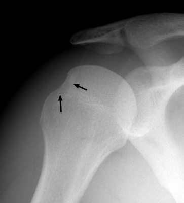
Figure 1 illustrates the classic plain radiographic manifestation of a Hill-Sachs defect on an anteroposterior projection obtained in maximal internal rotation. Rotating the humerus internally profiles the posterolateral margin of the humeral head tangentially against the radiographic cassette. The lesion manifests as a pronounced focal flattening and vertical cortical condensation line along the superior posterolateral articular margin. This appearance results from permanent compressive collapse of the subchondral trabecular bone following acute anterior dislocation over the rigid anterior-inferior glenoid rim.
4. Pathophysiology & Biomechanical Mechanism
The glenohumeral joint represents an inherently mobile ball-and-socket articulation characterized by minimal intrinsic bony constraint. The shallow glenoid fossa accommodates only approximately one-third of the hemispherical humeral head at any point during physiologic range of motion. Joint stability relies heavily on dynamic muscular forces (the rotator cuff force couple) and static capsulolabral restraints, particularly the anterior-inferior glenohumeral ligament (AIGHL) complex and the fibrocartilaginous glenoid labrum.
Anterior glenohumeral dislocation typically occurs when an indirect traumatic force drives the arm into hyperabduction, external rotation, and horizontal extension (the cocking position). As the humeral head translates anteriorly and inferiorly, the static anterior-inferior stabilizers undergo tensile failure, resulting in an avulsion of the anterior labrum (Bankart lesion), an anterior labroligamentous periosteal sleeve avulsion (ALPSA), or a capsular rupture. Once the humeral head breaches the anterior glenoid rim, it translates across the osseous margin of the scapula.
The biomechanical creation of the Hill-Sachs defect stems from a fundamental mismatch in the material properties of the two opposing osseous structures:
- Cortical and Trabecular Density Disparity: The anterior-inferior rim of the glenoid is constructed of dense, highly mineralized cortical bone supported by an adjacent subchondral plate designed to withstand repetitive compressive and shear loads. Conversely, the posterolateral superior aspect of the humeral head consists of a thin cortical shell (0.5 to 1.5 mm in thickness) enclosing porous, compliant cancellous trabecular bone.
- Compaction Kinetics: When the humeral head displaces anteriorly, its posterolateral margin is wedged forcefully against the sharp, hard anterior glenoid rim. The applied load exceeds the compressive yield strength of the cancellous bone, driving the trabecular arches past their elastic limit into plastic deformation, microfracture, and permanent structural compaction.
- Irreversible Osseous Loss: Because compacted trabecular bone possesses no elastic recoil, the resulting defect creates a permanent grooved or wedge-shaped excavation. Over time, fibrocartilaginous metaplasia and scar tissue form at the base of the crater, but the missing osseous volume is never physiologically reconstituted.
In subsequent functional motion, this structural defect alters joint kinematics. During arm abduction and external rotation, the posterolateral humeral head rotates anteriorly toward the glenoid face. If the defect is wide or deep enough to extend beyond the functional contact area of the glenoid, its medial edge can drop over and catch against the anterior glenoid rim (dynamic engagement), levering the humeral head anteriorly and precipitating recurrent dislocation under low-energy loads.
5. Modality-Specific Imaging Findings
Plain Radiography
Plain radiography represents the initial diagnostic modality in evaluating acute shoulder trauma and suspected instability. However, standard projections performed in external rotation or neutral alignment frequently miss the lesion, as the greater tuberosity rotates into profile and obscures the posterolateral defect behind its dense cortical outline:
- Anteroposterior (AP) Internal Rotation View: The humerus is placed in 60 to 80 degrees of internal rotation. This projection brings the posterolateral superior contour of the humeral head into tangential profile against the cassette. The defect appears as a vertical, linear radiolucent groove bordered by a sclerotic margin (“line of condensation”) or an abrupt loss of circular articular sphericity (“hatchet deformity”).
- Stryker Notch View: The patient is positioned supine with the ipsilateral hand resting on top of the head and the elbow pointing anteriorly. The X-ray beam is angled 10 degrees cephalad and centered directly over the coracoid process. This projection profiles the posterosuperior and posterolateral margin of the humeral head unobstructed by the acromion process, delivering the highest plain radiographic sensitivity (88% to 92%) for diagnosing and estimating the depth of a Hill-Sachs lesion.
- Didiee View: The patient is placed prone with the hand resting on the ipsilateral iliac crest and the thumb pointing cephalad (elbow abducted 45 degrees). The beam is aimed 45 degrees laterally toward the glenohumeral joint. This projection provides an unobstructed view of the posterolateral humeral head while simultaneously profiling the anterior-inferior glenoid rim to detect concomitant bony Bankart fractures.
- Garth (Apical Oblique) View: The patient is seated and rotated 45 degrees toward the affected shoulder, with the beam directed 45 degrees caudad through the glenohumeral joint. The Garth view is especially valuable in acute trauma because it visualizes both anterior-inferior glenoid rim fractures and posterior-superior humeral head compression fractures on a single exposure without requiring painful internal or external rotation of the injured extremity.
Computed Tomography
Computed tomography (CT), particularly with multiplanar 2D reformats and 3D volume rendering, represents the gold standard for defining osseous morphology and quantifying bone loss:
- Axial 2D CT: Identifies the precise depth, transverse width, and craniocaudal extent of the wedge-shaped compaction defect. The lesion is characteristically situated within the proximal 20 mm of the humeral head, at or above the level of the coracoid process tip. On axial slices, the normal circular arc of the humeral articular surface is abruptly flattened or excavated posterolaterally.
- 3D Volume Rendering with Digital Subtraction: Digital subtraction of the scapula generates a clear, unobstructed 3D model of the humeral head. This allows direct inspection of the defect’s orientation, width, and surface area relative to the rotator cuff footprint.
- Bipolar Bone Loss Quantification: CT provides the requisite metric precision for assessing bipolar bone defects. Using sagittal-oblique reformats aligned with the glenoid face, the inferior glenoid circle method (Pico technique) determines the expected diameter (D) and the missing anterior bone width (d). Concurrent measurement of the Hill-Sachs interval (HSI) establishes whether the lesion is “on-track” or “off-track”.
Magnetic Resonance Imaging & Ultrasound
Magnetic resonance imaging (MRI) and MR arthrography offer comprehensive assessment of both osseous defects and concomitant soft-tissue pathology:
- Bone Marrow Edema Patterns: In acute and subacute settings, fat-suppressed T2-weighted and proton density (PDFS) sequences demonstrate extensive hyperintense marrow edema within the posterolateral trabecular bone underlying the defect. This high signal reflects trabecular microfractures and hemorrhage, confirming recent impaction even when cortical depression is subtle.
- Chondral Integrity & Signal Voids: T1-weighted sequences delineate the cortical notch as a low-signal defect. Gradient echo (T2*) and fluid-sensitive sequences visualize hyaline cartilage fibrillation, cartilage delamination, and subchondral plate disruption, distinguishing purely cartilaginous impactions from deep osteochondral fractures.
- Associated Soft-Tissue Pathology: MRI excels at diagnosing concomitant anterior capsulolabral disruptions (fibrous Bankart, ALPSA, Perthes, GLAD), humeral avulsions of the glenohumeral ligament (HAGL), and superior labral anterior-to-posterior (SLAP) tears. In patients older than 40 years presenting with traumatic anterior dislocation, MRI is mandatory to evaluate for concurrent full-thickness rotator cuff tears (supraspinatus and subscapularis).
- High-Resolution Ultrasound: Utilizing a high-frequency linear transducer placed over the posterior glenohumeral joint with the arm in internal rotation, sonography depicts the defect as an abrupt focal step-off or discontinuity in the hyperechoic humeral cortical contour. While ultrasound cannot reliably quantify volumetric bone loss, it serves as an accessible bedside tool for rapid triage in emergency settings.
6. Diagnostic Utility, Sensitivity & Specificity
The diagnostic performance of imaging modalities for identifying Hill-Sachs lesions varies substantially based on technical projections and spatial resolution:
- Plain Radiographic Projections: Standard AP views in external rotation demonstrate poor sensitivity (approximately 50%), frequently missing significant defects. AP views in internal rotation improve sensitivity to 65% to 80%. The Stryker notch view achieves the highest plain film sensitivity, ranging from 88% to 92%, with a specificity exceeding 95%. The Didiee view provides 80% to 85% sensitivity and high specificity for anterior-inferior rim evaluation.
- Computed Tomography: Multidetector helical CT demonstrates 96% to 100% sensitivity and 98% to 100% specificity for detecting cortical breaches and cancellous compaction. CT is considered the imaging gold standard for linear and volumetric quantification of osseous defects. Inter-observer and intra-observer reliability for CT-based glenoid track and Hill-Sachs interval measurements yield intraclass correlation coefficients (ICC) between 0.78 and 0.88.
- Magnetic Resonance Imaging: Non-contrast 1.5T and 3T MRI demonstrate 90% to 95% sensitivity and 95% to 98% specificity for osseous Hill-Sachs lesions. MR arthrography increases sensitivity to 95% to 97%, particularly for superficial chondral impactions and subtle subchondral fractures.
- Prevalence Patterns: A Hill-Sachs defect is identified in 65% to 70% of primary acute anterior shoulder dislocations and in 80% to 100% of cases involving recurrent anterior instability. The presence of a large or deep Hill-Sachs lesion on initial imaging correlates strongly with recurrent instability episodes.
7. Differential Diagnosis & Radiographic Mimics
Accurate interpretation requires distinguishing the pathologic Hill-Sachs compression fracture from benign anatomic variants and other destructive humeral head conditions:
| Sign / Condition | Distinguishing Radiographic Features | Clinical Presentation | Key Differentiating Test |
|---|---|---|---|
| Hill-Sachs Sign (Classic Anterior Impaction) | Wedge-shaped cortical depression along the superior posterolateral humeral head within the proximal 20 mm; accompanied by a vertical line of condensation | History of acute or recurrent anterior glenohumeral dislocation; anterior apprehension and relocation tests positive | Stryker notch radiograph or axial CT; fat-suppressed MRI demonstrates bone marrow edema in acute phase |
| Normal Anatomical Groove (“Bare Area” of Humeral Head) | Smooth, physiologic cortical concavity located on the posterolateral humeral head, but situated more than 20 to 25 mm inferior to the superior articular margin | Asymptomatic, normal shoulder biomechanics; incidental radiographic finding without instability history | Axial CT or MRI; demonstrates intact overlying cortical bone, absence of trabecular compaction, and lack of bone marrow edema |
| Reverse Hill-Sachs (McLaughlin Lesion) | Vertical impaction fracture located on the anteromedial aspect of the humeral head, resulting from impaction against the posterior glenoid rim | History of posterior shoulder dislocation (seizure, electrocution, direct trauma); arm locked in internal rotation | Axial CT or AP radiograph with 45-degree external rotation; reveals anteromedial subchondral depression |
| Hatchet Deformity (Ankylosing Spondylitis) | Severe erosive scalloping and extensive bone loss of the superolateral humeral head, creating an exaggerated hatchet appearance | Bilateral shoulder pain, stiffness, inflammatory axial spine pain, sacroiliitis, HLA-B27 positivity | Pelvic/spinal radiographs showing sacroiliitis; serology; MRI demonstrates diffuse synovial pannus without dislocation history |
| Avascular Necrosis (Cruess / Ficat Stage II-III) | Subchondral radiolucent line (“crescent sign”) followed by segmental collapse of the superior articular dome; mottled sclerosis | Insidious, progressive mechanical shoulder pain; risk factors include high-dose corticosteroids, sickle cell disease, alcoholism | Coronal and axial MRI; demonstrates pathognomonic geographic “double-line sign” on T2-weighted sequences; lesion centered anterosuperiorly |
| Subchondral Degenerative Geode | Well-defined, rounded cystic radiolucency with a sclerotic rim located beneath the articular surface; associated with joint space narrowing and osteophytes | Chronic, progressive shoulder pain, crepitus, and stiffness in an older individual; primary or secondary osteoarthritis | Standard AP and axillary radiographs; demonstrates classic triad of osteoarthritis (narrowing, subchondral sclerosis, osteophytes) |
8. Clinical Significance & Management Implications
The clinical management of anterior shoulder instability has evolved from focusing solely on anterior soft-tissue repairs to comprehensively evaluating bipolar bone loss. Seminal biomechanical investigations by Burkhart, De Beer, Yamamoto, Itoi, and Di Giacomo established that isolated soft-tissue Bankart repairs fail in up to 67% of patients when significant osseous defects are present. The interaction between anterior glenoid bone loss and humeral head bone loss dictates the risk of postoperative recurrence.
The modern surgical paradigm relies on the concept of the Glenoid Track and the Hill-Sachs Interval:
- The Glenoid Track (GT): Biomechanical studies demonstrate that as the arm moves through 90 degrees of abduction and progressive external rotation (the functional ABER position), the contact zone between the glenoid and the humeral head sweeps from the inferomedial to the superolateral aspect of the humeral head. In an intact shoulder, the width of this contact zone (glenoid track) equals approximately 84% of the total inferior glenoid diameter:
GT = 0.84 × D(where D is the diameter of the inferior glenoid circle). - Correction for Glenoid Bone Loss: When an anterior glenoid bone defect of width d is present, the effective glenoid track width is shortened accordingly:
GT = 0.84 × (D − d). - The Hill-Sachs Interval (HSI): The HSI represents the total medial-to-lateral extent of humeral bone loss, measured from the medial articular margin of the rotator cuff (infraspinatus) footprint to the most medial edge of the Hill-Sachs defect. It is calculated as:
HSI = HS + BB(where HS is the width of the Hill-Sachs defect, and BB is the width of the intact bone bridge between the cuff footprint and the lateral border of the defect). - On-Track vs Off-Track Categorization:
• On-Track Lesion (HSI ≤ GT): The entire Hill-Sachs defect stays within the glenoid track during functional range of motion; the lesion does not engage the anterior glenoid rim.
• Off-Track Lesion (HSI > GT): The medial border of the Hill-Sachs defect extends medial to the glenoid track. In abduction and external rotation, the defect catches and engages over the anterior glenoid rim, levering the humeral head anteriorly.
This biomechanical categorization guides an evidence-based surgical decision algorithm:
- Glenoid Bone Loss < 13.5% (or < 20%) AND On-Track Hill-Sachs: Isolated arthroscopic Bankart repair with capsulolabral reconstruction restores stability, yielding low recurrence rates (< 5% to 10%).
- Glenoid Bone Loss < 13.5% (or < 20%) AND Off-Track Hill-Sachs: Performing an isolated Bankart repair carries a failure and redislocation rate of 30% to 50%. The recommended treatment is an Arthroscopic Bankart Repair combined with Remplissage (infraspinatus tenodesis and posterior capsulodesis into the Hill-Sachs crater). By filling the defect with vascularized soft tissue, the intra-articular defect is converted into an extra-articular footprint, preventing dynamic engagement. Recent meta-analyses confirm higher return to sport and significantly lower revision rates with this combined procedure.
- Subcritical or Critical Glenoid Bone Loss (≥ 13.5% to 25%) regardless of Track Status, or Off-Track with Borderline Glenoid Loss: Soft-tissue procedures fail to reliably restore stability. The gold standard is a Latarjet Coracoid Transfer (or iliac crest / distal tibial allograft reconstruction). Transferring the coracoid process with its conjoined tendon to the anterior-inferior glenoid neck restores the glenoid diameter (D), expanding the glenoid track and converting an off-track lesion to an on-track configuration (the triple-blocking effect: osseous augmentation, dynamic conjoint tendon sling, and capsular repair).
- Massive Humeral Head Defects (> 30% to 40% of articular surface): In rare cases of massive segmental humeral bone loss, soft-tissue tenodesis is insufficient: structural osteoarticular allograft reconstruction, partial humeral resurfacing, or shoulder arthroplasty is indicated.
9. Master Class Pro-Tip
Master Class Pro-Tip: Always measure the Hill-Sachs Interval (HSI) from the medial articular margin of the infraspinatus footprint rather than the lateral cortical border of the greater tuberosity. Neglecting the intact bone bridge (BB) leads to systematic underestimation of the true glenoid track engagement risk. Intraoperatively, during diagnostic arthroscopy, place the arm in 90 degrees of abduction and progressive external rotation while viewing from the anterosuperior portal. If the posterolateral humeral head defect slips over the anterior glenoid rim with an audible or palpable clunk, the lesion is dynamically engaging and an isolated Bankart repair will fail: perform an immediate arthroscopic remplissage or convert to a Latarjet procedure.
Master Class Radiological Pearl: Humeral Head Bone Loss and Dynamic Engagement
10. Evidence-Based Synthesis
| Author & Year | Study Design | Sample Size | Key Findings & Diagnostic Accuracy | Clinical Implication | Level of Evidence | DOI |
|---|---|---|---|---|---|---|
| Hill & Sachs (1940) | Seminal Radiographic Case Series | N = 119 | Identified posterolateral humeral head grooved defects in 74% of recurrent dislocations and 27% of acute primary dislocations; established AP internal rotation as standard projection | Seminal description establishing posterolateral impaction fracture as pathognomonic consequence of anterior dislocation | Level IV | DOI |
| Burkhart & De Beer (2000) | Prospective Clinical Cohort Study | N = 194 | Reported 67% recurrence after arthroscopic Bankart repair in patients with significant bone defects (inverted-pear glenoid or engaging Hill-Sachs) compared to 4% in those without osseous defects | Identified bone loss as the primary etiology of arthroscopic stabilization failure; established concept of engaging vs non-engaging lesions | Level IV | DOI |
| Yamamoto et al. (2007) | Biomechanical Cadaveric & Anatomic Investigation | N = 14 cadaveric shoulders | Defined the glenoid track as the contact zone between glenoid and humeral head during abduction and external rotation; width measured 84% ± 14% of glenoid width | Established scientific foundation for modern dynamic engagement assessment and bipolar bone loss quantification | Level V | DOI |
| Provencher et al. (2012) | Comprehensive Instructional Review | N = Narrative / Multi-study synthesis | Comprehensive synthesis of diagnostic imaging modalities, classification systems, and surgical management thresholds for humeral bone loss | Recommends cross-sectional CT/MRI volumetric quantification and provides guidelines for remplissage vs bone grafting | Level V | DOI |
| Di Giacomo, Itoi, & Burkhart (2014) | Expert Biomechanical Consensus & Algorithm | N = Biomechanical / Mathematical consensus | Developed the quantitative On-Track / Off-Track classification paradigm combining glenoid rim bone loss and Hill-Sachs interval: HSI = HS + BB vs GT = 0.84*(D – d) | Transformed clinical decision-making from subjective arthroscopic assessment to objective, preoperative imaging-based algorithmic selection | Level V | DOI |
| Masud et al. (2023) | Systematic Review / Bayesian Network Meta-Analysis | N = 3,467 shoulders (39 studies) | Network meta-analysis comparing Bankart repair, Bankart + remplissage, and Latarjet; bone-addressing procedures had significantly lower recurrence than Bankart alone | Demonstrates superior stability and return to sport when humeral or glenoid bone defects are addressed operatively | Level I | DOI |
| Ahmed et al. (2024) | Systematic Review / Meta-Analysis | N = 1,027 patients (15 comparative studies) | Bankart repair with remplissage yielded significantly lower revision rates (OR = 0.28, p < 0.001) and higher return to pre-injury sports compared to Bankart repair alone | Validates remplissage as the gold standard adjunct for off-track Hill-Sachs lesions with subcritical glenoid bone loss | Level I | DOI |
| Bulleit et al. (2024) | Systematic Review / Meta-Analysis | N = 4,812 patients (46 studies) | Systematic analysis identifying off-track Hill-Sachs lesions, glenoid bone loss > 15%, and young age as the primary independent predictors of Bankart failure | Confirms that preoperative calculation of the glenoid track and Hill-Sachs interval is mandatory prior to soft-tissue stabilization | Level I | DOI |
11. References
- Hill HA, Sachs MD. The grooved defect of the humeral head: A frequently unrecognized complication of dislocation of the shoulder joint. Radiology. 1940;35(6):690-700. DOI [Level IV: Seminal Radiographic Case Series]
- Burkhart SS, De Beer JF. Traumatic glenohumeral bone defects and their relationship to failure of arthroscopic Bankart repairs: significance of the inverted-pear glenoid and the engaging Hill-Sachs lesion. Arthroscopy. 2000;16(7):677-694. DOI [Level IV: Prospective Clinical Cohort Study]
- Yamamoto N, Itoi E, Abe H, et al. Contact between the glenoid and the humeral head in abduction, external rotation, and horizontal extension: A new concept of glenoid track. J Shoulder Elbow Surg. 2007;16(5):649-656. DOI [Level V: Biomechanical Cadaveric & Anatomic Investigation]
- Provencher MT, Frank RM, Leclere LE, et al. The Hill-Sachs Lesion: Diagnosis, Classification, and Management. J Am Acad Orthop Surg. 2012;20(4):242-252. DOI [Level V: Comprehensive Instructional Review]
- Di Giacomo G, Itoi E, Burkhart SS. Evolving Concept of Bipolar Bone Loss and the Hill-Sachs Lesion: From ‘Engaging/Non-Engaging’ Lesion to ‘On-Track/Off-Track’ Lesion. Arthroscopy. 2014;30(1):90-98. DOI [Level V: Expert Biomechanical Consensus & Algorithm]
- Masud S, Momtaz D, Betsch M, et al. A comprehensive comparison and evaluation of surgical techniques for anterior shoulder instability: a Bayesian network meta-analysis. J Shoulder Elbow Surg. 2023;33(2):474-486. DOI [Level I: Systematic Review / Meta-Analysis]
- Ahmed AF, Polisetty TS, Wang C, et al. Higher return to sport and lower revision rates when performing arthroscopic Bankart repair with remplissage for anterior shoulder instability with a Hill-Sachs lesion: a meta-analysis. J Shoulder Elbow Surg. 2024;33(7):1621-1632. DOI [Level I: Systematic Review / Meta-Analysis]
- Bulleit CH, Hurley ET, Jing C, et al. Risk factors for recurrence following arthroscopic Bankart repair: a systematic review. J Shoulder Elbow Surg. 2024;33(10):2375-2384. DOI [Level I: Systematic Review / Meta-Analysis]
Last Updated on September 28, 2026
🔗 Knowledge Graph & Clinical Correlates
OrthopaedicsOne Living Semantic Network🛠️ Operative Techniques
- Turf Toe Injuries Sports Medicine
- Total Shoulder Arthroplasty for Glenohumeral Arthritis with an Intact Rotator Cuff Shoulder and Elbow
- The Role of Tendon Transfers in Massive Rotator Cuff Tears Shoulder and Elbow
- The Disabled Throwing Shoulder Sports Medicine
🩺 Clinical Conditions & Indications
- Turf toe Introduction Despite its rather benign namesake, turf toe is a incapacitating injury that plagues many athletes who participate...
- Therapeutic exercise for the shoulder Therapeutic exercise for the shoulder Range of Motion and Flexibility Pendulums Table Slides into flexion, external rotation, and...
- The cross-sectional study 1. High-Yield Summary Fundamental Architecture: A cross-sectional study (prevalence study or transverse survey) is an observational epidemiological design...
- Subacromial impingement 1. High-Yield Summary Subacromial impingement, now increasingly designated subacromial pain syndrome (SAPS) or rotator cuff-related shoulder pain (RCRSP),...
🔬 Anatomy, Pathology & Diagnostics
- Trough Line Glenohumeral Joint & Rotator Cuff
- The Role of Periosteum in Cartilage Repair General Musculoskeletal
- The morphology of the acromion according to Bigliani Glenohumeral Joint & Rotator Cuff
- The Biomechanical Case for Labral Débridement General Musculoskeletal