
1. High-Yield Overview & Pathognomonic Essence
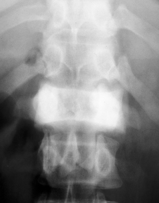
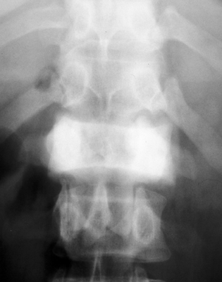
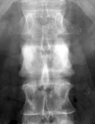
- Fundamental Radiographic Appearance: The ivory vertebra sign denotes a solitary (or infrequently multiple) vertebral body exhibiting marked, homogeneous, diffuse radiodensity and sclerosis, presenting as an intense “chalky white” or “ivory” appearance that contrasts sharply against adjacent normal or osteopenic vertebral bodies on plain radiographs and computed tomography, with characteristic initial preservation of vertebral contours and adjacent intervertebral disc heights.
- Pathognomonic Musculoskeletal Conditions: The sign is the classic radiographic manifestation of diffuse cancellous and cortical hyperostosis representing a well-established diagnostic triad: osteoblastic skeletal metastasis (most frequently carcinoma of the prostate in men and breast in women), Paget disease of bone (osteitis deformans), and lymphoma (Hodgkin lymphoma and non-Hodgkin lymphoma). Less common etiologies include chronic tuberculous or pyogenic osteomyelitis, primary bone sarcomas (osteosarcoma, chordoma), and benign or metabolic disorders (sclerotic hemangioma, mastocytosis, fluorosis).
- Critical Diagnostic & Surgical Implications: Identification of an ivory vertebra is an immediate clinical red flag mandating urgent systemic oncologic and metabolic staging. Anatomic morphologic analysis serves as the primary non-invasive discriminator: outward osseous expansion with trabecular coarsening confirms Paget disease; normal vertebral dimensions with pedicle erosion indicate osteoblastic metastasis; and anterior cortical erosion or scalloping indicates lymphomatous nodal erosion. In cases without known primary malignancy, percutaneous CT-guided transpedicular biopsy provides definitive histological verification, while the Spinal Instability Neoplastic Score (SINS) guides operative stabilization and decompression.
2. Diagnostic Sign Summary Matrix
| Primary Modality | Anatomic Location | Primary Pathology | Pathognomonic Morphology | Diagnostic Utility & Sensitivity |
|---|---|---|---|---|
| Plain Radiography (AP & Lateral) | Thoracic and lumbar spine (most common); cervical spine | Osteoblastic metastasis (prostate, breast), Paget disease of bone, or lymphoma | Uniform, dense, homogeneous radiopacity of a single vertebral body (“chalky white” vertebra); intact adjacent disc spaces | First-line screening; high conspicuity once 30% to 50% bone mineral density increases; distinguishes Paget expansion from neoplastic preservation of size |
| Computed Tomography (CT) | Vertebral body centrum, pedicles, laminae, and posterior elements | Dense trabecular hyperostosis, new bone formation, and cortical architecture alterations | Marked hyperattenuation (>600 to 1000 Hounsfield units) replacing normal cancellous trabeculae; clear delineation of cortical expansion versus cortical destruction | Gold standard for osseous matrix details, cortical integrity, posterior wall breach, and CT-guided transpedicular biopsy trajectory planning; 100% spatial resolution |
| Magnetic Resonance Imaging (MRI) | Vertebral marrow space, spinal canal, neural foramina, and paravertebral soft tissues | Replacement of marrow fat by tumor stroma, dense woven bone, or fibrous tissue | Profound low signal intensity (hypointense signal void) on T1- and T2-weighted sequences; STIR/T2 hyperintensity in active cellular tumor zones and epidural mass | Gold standard for staging spinal cord or cauda equina compression; differentiates retained fatty marrow islands in Paget disease from diffuse tumor replacement |
| Nuclear Scintigraphy & PET-CT | Total axial and appendicular skeleton; systemic lymph nodes and visceral organs | Accelerated osteoblastic turnover, skeletal metabolic activity, or hypermetabolism | Intense focal radiotracer uptake on 99mTc-MDP bone scintigraphy; elevated SUVmax on 18F-FDG PET-CT (lymphoma, metastasis) or 68Ga-PSMA PET-CT (prostate) | High sensitivity for identifying occult polyostotic disease, skip skeletal metastases, or multi-station lymphoma; essential for whole-body staging and therapy monitoring |
3. Clinical Case Imaging & Standardized Figures
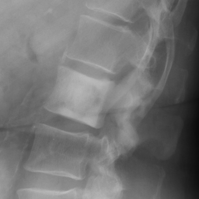
The standardized radiographic series below illustrates the classical presentations of the ivory vertebra sign across its primary differential diagnoses, contrasting the expansile, trabeculated hyperostosis of skeletal Paget disease with the dense, non-expansile osteoblastic infiltration of metastatic carcinoma:
4. Pathophysiology & Biomechanical Mechanism
The pathophysiological foundation of the ivory vertebra sign lies in the profound alteration of physiological bone remodeling, where endosteal and periosteal new bone deposition drastically outpaces osteoclastic bone resorption. Although the end-stage radiographic appearance of intense radiopacity is shared across disparate disease entities, the underlying cellular, biochemical, and molecular pathways diverge significantly among the primary diagnostic categories:
In osteoblastic skeletal metastases (predominantly prostate adenocarcinoma in men and breast adenocarcinoma in women), malignant tumor cells colonize the bone marrow microenvironment via Batson’s valveless vertebral venous plexus. Once established in the marrow cavity, tumor cells initiate complex paracrine signaling networks with resident osteoblasts and stromal cells. Prostate and breast cancer cells secrete high levels of endothelin-1 (ET-1), transforming growth factor-beta (TGF-beta), bone morphogenetic proteins (BMPs), platelet-derived growth factor (PDGF), and Wnt pathway agonists. ET-1 binds with high affinity to endothelin A (ET_A) receptors on osteoprogenitor cells, suppressing Dickkopf-1 (DKK-1) expression and releasing the canonical Wnt/beta-catenin signaling cascade from inhibition. This molecular uncoupling drives exuberant osteoblast proliferation and differentiation. The activated osteoblasts rapidly deposit vast quantities of unmineralized osteoid matrix in a disorganized woven pattern. Subsequent hypermineralization of this woven bone completely obliterates the intertrabecular marrow spaces, transforming the sponge-like cancellous vertebral body into a solid block of hyperdense bone.
In Paget disease of bone (osteitis deformans), the primary disturbance is a disorder of osteoclast biology, frequently linked to genetic mutations in SQSTM1 (encoding the p62 ubiquitin-binding protein) and heightened sensitivity to receptor activator of nuclear factor kappa-B ligand (RANKL). Following an initial hyperactive osteolytic phase, the disease transitions into an intense, chaotic osteoblastic sclerotic phase. Osteoblasts attempt to repair the extensive osteoclastic resorption by laying down large volumes of new bone at an accelerated rate. Because this process is uncoordinated and rapid, the newly synthesized bone consists of haphazard, irregular lamellar units separated by prominent cement lines, producing the classic “mosaic” or “jigsaw puzzle” histological appearance. Appositional periosteal and endosteal bone formation leads to true osseous hypertrophy, causing circumferential enlargement of the vertebral body and marked cortical thickening.
In lymphoma (both Hodgkin lymphoma and non-Hodgkin lymphoma), marrow infiltration by neoplastic lymphoid cells triggers an intense host desmoplastic and osteosclerotic stromal reaction mediated by localized cytokine release, including interleukin-6 (IL-6), tumor necrosis factor-alpha (TNF-alpha), and lymphotoxin. In addition to intraosseous sclerosis, contiguous retroperitoneal and paravertebral lymphadenopathy frequently exerts direct pulsatile extrinsic mechanical pressure against the anterior vertebral body. Because the anterior longitudinal ligament is firmly anchored to the superior and inferior vertebral endplates but loosely adherent across the mid-vertebral waist, chronic pressure erosion from enlarged nodes produces focal anterior cortical scalloping, historically termed the “gallows” sign.
From a biomechanical perspective, the profound mineral density of an ivory vertebra creates an illusion of structural strength. In reality, sclerotic bone exhibits substantial biomechanical vulnerability. Disorganized woven bone in metastatic carcinoma and mosaic lamellar bone in Paget disease have an abnormal modulus of elasticity, impaired ductility, and increased brittleness. Under physiological cyclical axial loading and shear stress, the hyperdense bone cannot dissipate energy through normal microstructural deformation. In Paget disease, this brittleness predisposes to transverse insufficiency fractures (“chalk-stick” or “banana-type” fractures). In metastatic malignancy, the underlying malignant infiltration compromises trabecular architecture, rendering the vertebral body susceptible to acute pathological compression or burst fractures with posterior wall retropulsion into the spinal canal.
5. Modality-Specific Imaging Findings
Plain Radiography
Standard anteroposterior (AP) and lateral radiographs remain the foundational imaging examination for detecting the ivory vertebra sign. The classic presentation is an isolated vertebral body exhibiting uniform, chalky white radiopacity that stands out boldly against adjacent normal or osteopenic vertebrae. Critical morphological features must be evaluated systematically on plain films:
- Vertebral Body Dimensions: Assessing vertebral size is the most decisive plain radiographic step. Enlargement of the vertebral body in both anteroposterior and transverse dimensions with thickened, hyperdense peripheral cortical margins (“picture frame” vertebra) is pathognomonic for Paget disease of bone. In contrast, osteoblastic metastases and lymphoma characteristically preserve normal vertebral body dimensions without osseous expansion.
- Posterior Element Architecture: Sclerosis or destruction of the pedicles and posterior elements must be scrutinized on AP projections. Osteoblastic metastases frequently involve the pedicles; cortical erosion or absent pedicle outlines (the “winking owl” sign when unilateral, or “blind vertebra” when bilateral) strongly supports metastatic malignancy. In Paget disease, the pedicles and neural arch may be sclerotic and thickened, but cortical continuity is maintained without destructive osteolysis.
- Anterior Cortical Contour: On lateral radiographs, smooth concave erosion or scalloping of the anterior vertebral cortex in the presence of intact endplates is highly characteristic of Hodgkin lymphoma, resulting from extrinsic erosion by adjacent retroperitoneal lymphadenopathy.
- Intervertebral Disc Space: In neoplastic etiologies and Paget disease, the adjacent intervertebral disc spaces remain strictly preserved in height without endplate erosions. Immediate narrowing or destruction of the adjacent disc space combined with endplate irregularities firmly excludes uncomplicated neoplasia and points to infectious spondylodiscitis (such as tuberculous Pott disease or chronic pyogenic osteomyelitis).
Computed Tomography
Multi-detector computed tomography (CT) with multiplanar 2D reformations (axial, coronal, sagittal) and bone-algorithm reconstruction windows provides unmatched spatial resolution of the cortical and trabecular architecture. CT confirms the diagnosis by demonstrating diffuse, extreme hyperattenuation within the vertebral body, where Hounsfield unit (HU) measurements routinely exceed 600 to 1200 HU, compared to normal cancellous bone values of 150 to 250 HU.
CT readily unmasks subtle distinguishing diagnostic features that may remain occult on plain films. In Paget disease, axial and sagittal CT views exquisitely demonstrate cortical expansion, endosteal cortical thickening, and coarsened, thickened vertical trabeculae. In osteoblastic metastases, CT reveals multiple confluent, round-to-oval osteosclerotic nodules, cortical disruption, and involvement of the pedicles, lamina, and spinous processes. In lymphoma, CT confirms anterior cortical erosion while demonstrating the causative prevertebral or retroperitoneal lymph node conglomerate encasing adjacent vascular structures. Crucially, CT evaluates the structural integrity of the posterior vertebral cortex and quantitatively assesses spinal canal stenosis resulting from retropulsed bone fragments or concentric pagetic expansion.
Magnetic Resonance Imaging & Ultrasound
Magnetic resonance imaging (MRI) is essential for staging neural axis compromise, characterizing bone marrow cellularity, and identifying soft-tissue extensions. On all conventional pulse sequences (T1-weighted, T2-weighted, and proton density), dense sclerotic bone produces a profound signal void, manifesting as marked hypointensity. However, fluid-sensitive sequences (T2-weighted fat-suppressed and STIR) and post-contrast T1-weighted images provide vital discriminatory information:
In metastatic carcinoma and lymphoma, the remaining marrow cavity and adjacent cortical breaches show high STIR and T2 signal corresponding to hypercellular tumor tissue, edema, and hypervascularity. Contrast-enhanced T1 sequences demonstrate prominent enhancement within epidural soft-tissue components, precisely mapping the degree of thecal sac indentation, spinal cord displacement, and neural foraminal stenosis. In Paget disease, MRI typically reveals preservation of interspersed foci of fatty marrow (high T1 signal) within the coarsened trabecular network, and substantial epidural or paraspinal soft-tissue masses are distinctly absent unless rare secondary sarcomatous transformation (pagetic osteosarcoma) has supervened.
High-resolution musculoskeletal ultrasound has a negligible role in imaging the deep cancellous architecture of the vertebral body due to complete reflection of acoustic beams by overlying bony cortices. Nevertheless, point-of-care ultrasound serves as an indispensable adjunct for rapid evaluation and percutaneous needle biopsy of superficial primary malignancies (such as breast masses or thyroid nodules) and suspicious peripheral lymphadenopathy (cervical, supraclavicular, axillary, or inguinal stations), which frequently unlocks the primary histological diagnosis in patients presenting with an occult ivory vertebra.
6. Diagnostic Utility, Sensitivity & Specificity
The diagnostic sensitivity of plain radiography for early bone marrow infiltration is relatively low, as approximately 30% to 50% of trabecular bone mineral must be altered before osteosclerosis or osteolysis becomes clearly discernible to the human eye on conventional X-rays. Consequently, early metastatic colonization or early lymphomatous marrow infiltration may escape detection on initial screening radiographs. However, once the pathology has progressed to produce the fully developed ivory vertebra sign, radiographic conspicuity approaches 100%.
In terms of clinical specificity, the ivory vertebra sign carries near-absolute specificity for significant underlying musculoskeletal pathology. It is never a benign anatomical variant. Across large historical and contemporary imaging cohorts (Dennis 1961, Santos 2025), the etiological breakdown in adult patients demonstrates a consistent epidemiological pattern:
- Osteoblastic Metastatic Disease: Accounts for 50% to 70% of all solitary ivory vertebrae in adult populations. Carcinoma of the prostate represents the predominant cause in men over 50 years of age, while metastatic breast adenocarcinoma represents the leading cause in women. Other osteoblastic primary tumors include neuroendocrine (carcinoid) tumors, transitional cell bladder carcinoma, and gastrointestinal/pancreatobiliary adenocarcinomas.
- Paget Disease of Bone: Accounts for 20% to 25% of cases, typically presenting in individuals older than 55 years, with a slight male predominance.
- Lymphoma (Hodgkin & Non-Hodgkin): Accounts for 10% to 15% of cases, frequently presenting in young adults (Hodgkin lymphoma, aged 20 to 40 years) or elderly patients (non-Hodgkin lymphoma).
- Atypical & Rare Conditions: Comprise less than 5% of cases, including chronic tuberculous spondylitis (Pott disease), primary osteosarcoma of the spine, chordoma, sclerotic hemangioma, systemic mastocytosis, and fluorosis.
The combination of cross-sectional CT and contrast-enhanced MRI yields excellent diagnostic accuracy in differentiating these entities prior to tissue biopsy. The presence of true osseous expansion combined with coarsened trabecular architecture provides a specificity exceeding 95% for Paget disease of bone. Conversely, the presence of normal vertebral dimensions with pedicle destruction or extraosseous epidural mass provides greater than 90% specificity for metastatic malignancy or lymphoma.
7. Differential Diagnosis & Radiographic Mimics
| Sign / Condition | Distinguishing Radiographic Features | Clinical Presentation | Key Differentiating Test |
|---|---|---|---|
| Osteoblastic Metastasis (Prostate, Breast, Carcinoid) | Uniform or nodular sclerosis; normal vertebral body dimensions; pedicle erosion or destruction (“winking owl” sign); preserved adjacent disc space | Older adults (>50 years); axial spine pain; constitutional symptoms; history of primary malignancy (prostate, breast) | Serum PSA, CA 15-3; CT/MRI staging; percutaneous CT-guided transpedicular biopsy; 99mTc-MDP bone scintigraphy |
| Paget Disease of Bone (Osteitis Deformans) | Osseous expansion (enlarged vertebral body); marked cortical thickening producing “picture frame” vertebra; coarsened vertical trabeculae | Patients >55 years; frequently asymptomatic; deep ache, spinal stenosis, or kyphosis; elevated serum alkaline phosphatase | Markedly elevated serum alkaline phosphatase with normal liver transaminases; absence of soft-tissue mass on CT/MRI |
| Lymphoma (Hodgkin & Non-Hodgkin) | Homogeneous sclerosis; normal vertebral dimensions; classic anterior cortical scalloping / erosion (“gallows” sign); preserved disc space | Young adults (Hodgkin, 20-40 years) or older adults (NHL); B-symptoms (fever, night sweats, weight loss), peripheral adenopathy | Contrast-enhanced CT/MRI demonstrating extensive retroperitoneal/paraspinal adenopathy; excisional peripheral node biopsy |
| Chronic Osteomyelitis / Tuberculous Spondylitis | Dense reactive sclerosis; severe intervertebral disc space narrowing; irregular endplate destruction; prevertebral / psoas abscess | Progressive back pain, night pain, fever, chills, night sweats; elevated inflammatory markers (ESR, CRP); endemic exposure | Contrast MRI showing fluid-filled disc space and rim-enhancing abscess; CT-guided aspiration for acid-fast bacilli and cultures |
| Primary Bone Sarcoma (Osteosarcoma / Chordoma) | Aggressive osteoid matrix mineralization; cortical destruction; large, calcified extraosseous soft-tissue mass; pedicle invasion | Adolescents or older adults; rapid onset of severe, unrelenting focal pain, palpable mass, early neurological deficits | Surgical or percutaneous core needle biopsy; staging CT/MRI; surgical oncology consultation for en bloc resection |
| Sclerotic Hemangioma / Systemic Mastocytosis | Prominent vertical trabecular thickening (“corduroy cloth” on plain films, “polka-dot” on axial CT); high T1/T2 fat signal | Typically incidental finding; mastocytosis presents with episodic flushing, pruritus, urticaria pigmentosa, diarrhea | MRI demonstrates characteristic high T1 and T2 intraosseous fat signal in hemangioma; serum tryptase and bone marrow biopsy for mastocytosis |
8. Clinical Significance & Management Implications
The discovery of an ivory vertebra sign on spinal imaging fundamentally directs the clinical and surgical management strategy. Because the vast majority of cases in adults represent metastatic malignancy, lymphoma, or active Paget disease, an organized, multidisciplinary diagnostic and therapeutic protocol is mandatory:
Systematic Diagnostic Protocol: Initial laboratory evaluation should include complete blood count, comprehensive metabolic panel, serum calcium, phosphate, intact parathyroid hormone, alkaline phosphatase (with bone-specific fraction), serum protein electrophoresis (SPEP) with immunofixation, serum free light chains, and dedicated tumor markers, notably serum prostate-specific antigen (PSA) in men and CA 15-3 and CEA in women. Whole-body staging should be executed promptly utilizing contrast-enhanced CT of the chest, abdomen, and pelvis, combined with 99mTc-MDP bone scintigraphy or 18F-FDG / 68Ga-PSMA PET-CT to evaluate systemic skeletal and visceral burden.
Percutaneous CT-Guided Biopsy: In patients without a verified oncologic history or whose imaging findings do not conclusively demonstrate the pathognomonic benign features of Paget disease (vertebral expansion with preserved fatty marrow), percutaneous CT-guided transpedicular core biopsy is the definitive diagnostic gold standard. Interventional proceduralists must be prepared for extreme bone hardness; standard hand-driven biopsy trocars frequently fail or deflect off the eburnated cortex, necessitating the use of motorized drill systems or heavy-duty trephine needles to acquire diagnostic marrow architecture and avoid non-diagnostic crush artifacts.
Spinal Stability & Surgical Indications: Mechanical stability must be formally evaluated using the Spinal Instability Neoplastic Score (SINS). Although dense blastic lesions score lower on the bone lesion quality parameter compared to lytic lesions, the brittleness of sclerotic woven bone predisposes to sudden insufficiency collapse under physiological mechanical load. Emergent surgical intervention (posterior decompression, separation surgery, and instrumented stabilization) is indicated in the setting of progressive neurological deficits, high-grade epidural spinal cord compression (ESCC scale grades 2 or 3), or mechanical spinal instability (SINS score of 13 to 18), followed by stereotactic body radiation therapy (SBRT).
Targeted Medical Therapy: Medical management is customized to the definitive histopathological diagnosis. In metastatic bone disease, systemic antiresorptive agents (intravenous zoledronic acid or subcutaneous denosumab) are administered to inhibit tumor-induced osteoclast recruitment, reduce skeletal-related events (SREs), and stabilize bone mineral turnover, alongside primary systemic systemic therapy (androgen deprivation therapy with novel antiandrogens for prostate cancer, or chemotherapy/HER2-targeted therapy for breast cancer). In symptomatic Paget disease, a single infusion of intravenous zoledronic acid (5 mg) provides rapid, long-lasting normalization of bone turnover, alleviation of bone pain, and prevention of progressive osseous deformity. In cases of painful, non-displaced pathological compression fractures without neurological compromise, percutaneous cement augmentation (vertebroplasty or kyphoplasty) may be considered, exercising caution regarding high cement injection pressures within densely sclerotic cancellous spaces.
9. Master Class Pro-Tip
Master Class Pro-Tip: When evaluating a solitary ivory vertebra, execute a disciplined three-step morphological checklist before requesting a biopsy: First, inspect vertebral dimensions on cross-sectional CT; true outward osseous expansion with cortical thickening and coarsened trabeculae is virtually pathognomonic for Paget disease of bone, eliminating the need for an invasive biopsy. Second, examine the anterior vertebral cortex and prevertebral space; sharp anterior cortical scalloping with preserved intervertebral disc spaces points directly toward adjacent lymphadenopathy from Hodgkin or non-Hodgkin lymphoma. Third, evaluate the pedicles and contiguous disc spaces; preserved normal vertebral contours with pedicle sclerosis or erosion in an older adult strongly signifies osteoblastic metastasis (prostate or breast adenocarcinoma), whereas disc space narrowing and irregular endplate destruction firmly identify infectious spondylodiscitis. If a biopsy is mandated for occult metastatic disease, inform the interventional radiologist in advance to utilize a motorized drill or high-gauge trephine needle, as hand-driven trocars routinely fail against hyperdense eburnated bone.
Master Class Radiological Pearl: Ivory Vertebra Differentiation
10. Evidence-Based Synthesis
| Author & Year | Study Design | Sample Size | Key Findings & Diagnostic Accuracy | Clinical Implication | Level of Evidence | DOI |
|---|---|---|---|---|---|---|
| Dennis JM (1961) | Seminal Radiographic Case Series & Morphologic Classification | N = 31 patients presenting with isolated solitary dense vertebral bodies | Dennis established the classic diagnostic triad for the solitary dense vertebral body: osteoblastic metastases (prostate, breast), Paget disease of bone, and lymphoma (Hodgkin and non-Hodgkin). Outlined key radiographic discriminators including vertebral expansion in Paget disease versus normal size or pedicle destruction in metastatic neoplasia. | Seminal landmark study establishing the structured radiological differential diagnosis and proving that skeletal metastasis accounts for the majority of adult cases. | Level IV | DOI |
| Graham TS (2005) | Landmark Radiological Review & Pathophysiology Monograph | Expert narrative monograph and radiological synthesis | Graham defined the ivory vertebra sign as an increase in opacity of a vertebral body that retains its size and contours, with no change in the adjacent intervertebral disks. Synthesized the pathophysiologic mechanisms: osteoblastic tumor stimulation (endothelin-1, TGF-beta) in metastasis, disorganized woven and lamellar bone deposition in Paget disease, and lymphomatous osteosclerosis. | Provides modern definitive imaging criteria and diagnostic roadmap distinguishing benign from malignant causes based on morphology and anterior cortex remodeling. | Level V | DOI |
| Braun RA et al. (2016) | Cross-Sectional Comparative Multimodality Imaging Review | N = 12 confirmed cases with CT, MRI, and histological confirmation | Braun and colleagues demonstrated that cross-sectional CT and MRI are critical in elucidating the underlying etiology of an ivory vertebra. MRI differentiates metastases and lymphoma (low T1 signal with replacement of marrow fat and associated soft-tissue or epidural components) from Paget disease (coarse trabeculae, preserved fatty marrow foci, cortical expansion) and infective spondylodiscitis (disc edema and fluid collections). | Demonstrated the indispensability of multimodality imaging (CT/MRI) over plain radiographs alone for precise non-invasive differentiation. | Level V | DOI |
| Matthews G, Dyer RB (2017) | Contemporary Radiological Imaging Review | Radiological sign review and diagnostic algorithm | Matthews and Dyer emphasized critical imaging nuances: anterior cortical scalloping points strongly toward lymphoma due to adjacent retroperitoneal adenopathy; enlarged vertebral dimensions and coarse trabecular thickening indicate Paget disease; and solitary or multiple hyperdense sclerotic foci without expansion in an older patient strongly indicate osteoblastic metastatic carcinoma. | Refines radiological discriminatory features on abdominopelvic and spinal CT, preventing misattribution and guiding targeted tissue sampling. | Level V | DOI |
| Tutar S et al. (2016) | Clinical Spine Imaging Case & Algorithmic Review | N = 1 index case with comprehensive spine surgical audit | Tutar and colleagues reported on an isolated ivory vertebra discovered during investigation of acute-on-chronic axial back pain. Outlined the clinical necessity of percutaneous CT-guided transpedicular biopsy when metastatic disease is suspected but no known primary tumor exists. | Validates the protocolized biopsy pathway for solitary sclerotic spinal lesions to achieve definitive histopathological diagnosis prior to oncologic or surgical therapy. | Level V | DOI |
| Liu SZ et al. (2020) | Clinical Diagnostic Case Report & Diagnostic Evaluation | N = 1 presentation of solitary sclerotic thoracic vertebra | Liu and colleagues demonstrated that an isolated ivory vertebra can be the inaugural presentation of occult metastatic prostate adenocarcinoma in the absence of urinary symptoms. Illustrated complete replacement of normal trabecular bone by dense sclerotic bone with elevated serum prostate-specific antigen (PSA). | Highlights the mandatory inclusion of serum tumor markers (PSA, CA 15-3, CEA) and whole-body bone scintigraphy in any male patient presenting with an ivory vertebra. | Level V | DOI |
| Pothiawala S (2022) | Emergency Department Cohort & Diagnostic Case Report | N = 1 acute emergency presentation with systemic review | Pothiawala identified the ivory vertebra sign on initial lumbar plain radiography in an emergency setting, leading to the rapid detection of metastatic adenocarcinoma from a primary gastric neoplasm. Discussed the diagnostic urgency of recognizing dense vertebral bodies in emergency back pain evaluations. | Reinforces the role of the ivory vertebra sign as an urgent red flag on emergency radiographs that mandates systemic staging and oncology referral. | Level V | DOI |
| Santos M et al. (2025) | Systematic Literature Review & Clinical Case Report | N = 1 index case; N = 42 pooled cases in systematic review | Santos and colleagues reviewed atypical and gastrointestinal origins of the ivory vertebra sign, identifying pancreatobiliary neoplasms as rare osteoblastic metastatic sources. Emphasized that 50% to 70% of ivory vertebrae in adults represent metastatic disease, with 20% to 25% Paget disease, and 10% to 15% lymphoma. | Provides contemporary epidemiological frequency distributions for ivory vertebra etiologies, underscoring the dominance of osteoblastic metastasis. | Level V | DOI |
| Krishnan S (2024) | Spine Surgical Review & Management Monograph | Comprehensive spine surgical review | Krishnan synthesized the surgical decision-making framework for patients with ivory vertebrae. Outlined protocols for structural stability assessment using the Spinal Instability Neoplastic Score (SINS), indications for cement augmentation (vertebroplasty/kyphoplasty) in pathological fractures, and surgical decompression for neural element compromise. | Integrates radiographic sign recognition into modern spine surgical workflows and stability scoring systems. | Level V | DOI |
11. References
- Dennis JM. The Solitary Dense Vertebral Body. Radiology. 1961;77(4):618-621. DOI [Level IV: Retrospective Case Series]
- Graham TS. The Ivory Vertebra Sign. Radiology. 2005;235(2):614-615. DOI [Level V: Expert Review]
- Braun RA, Milito CF, Goldman SM, Fernandes Ede Á. Ivory vertebra: imaging findings in different diagnoses. Radiol Bras. 2016;49(2):117-121. DOI [Level V: Pictorial Review]
- Matthews G, Dyer RB. The “ivory vertebra” sign. Abdom Radiol (NY). 2017;42(1):338-340. DOI [Level V: Radiological Review]
- Tutar S, Unal M, Yurtsever C, Ozturk E. Ivory vertebra sign. Spine J. 2016;16(7):e439-e440. DOI [Level V: Clinical Case Report]
- Liu SZ, Zhou X, Song A, Wang YP, Liu Y. The ivory vertebra sign. QJM. 2020;113(3):209-210. DOI [Level V: Clinical Vignette]
- Pothiawala S. A case report of ivory vertebra sign: an initial radiological manifestation of underlying abdominal malignancy. Clin Exp Emerg Med. 2022;9(4):359-362. DOI [Level V: Clinical Case Report]
- Santos M, Pinho M, Gouveia J, Sousa M, Peixoto L. Ivory Vertebra Phenomenon as a Sign of Metastatic Dissemination in Pancreatobiliary Neoplasia: A Case Report and Literature Review. Cureus. 2025;17(1):e77978. DOI [Level V: Literature Review]
- Krishnan S. Ivory Vertebra Sign. J Spinal Surg. 2024;11(1):38-40. DOI [Level V: Narrative Review]
Last Updated on September 28, 2026
🔗 Knowledge Graph & Clinical Correlates
OrthopaedicsOne Living Semantic Network🛠️ Operative Techniques
🩺 Clinical Conditions & Indications
- Torts 1. High-Yield Summary Medical tort liability in orthopaedic surgery arises when clinical care departs from accepted standards of...
- Timing of secondary sexual characteristics 1. High-Yield Overview & Clinical Purpose Biological Staging and Pubertal Timing: Secondary sexual characteristics, categorized via the Marshall...
- Thoracolumbar trauma Introduction Trauma to the lumbar or thoracic spine remains a clinical challenge from a diagnostic and management perspective....
- Thoracolumbar sprains 1. High-Yield Summary Definition & Core Distinction: Thoracolumbar sprains involve mechanical injury, micro-tearing, or partial disruption of the...
🔬 Anatomy, Pathology & Diagnostics
- Vertebroplasty and Kyphoplasty for Osteolytic Vertebral Collapse Lumbar Spine
- Vertebral endplate and marrow changes – Modic classification Lumbar Spine
- Vertebra prominens Surface Anatomy Lumbar Spine
- Tumbling Bullet Sign Lumbar Spine