
Contents
- Name of bone
- Location/Articulation
- Muscle and ligament attachments
- Surface anatomy
- Radiography
- Physical examination
- Embryology
- Anomalies
- Injuries/Disorders
Name of bone
Occipital
Location/Articulation
The occipital bone, a saucer-shaped membrane bone situated at the back and lower part of the cranium, is trapezoid in shape and curved on itself. It is pierced by a large oval aperture, the foramen magnum, through which the cranial cavity communicates with the vertebral canal.
- The curved, expanded plate behind the foramen magnum is named the squama occipitalis
- The thick, somewhat quadrilateral piece in front of the foramen is called the basilar part of occipital bone
- On either side of the foramen are the lateral parts of occipital bone
The occipital bone articulates with the two parietal bones, the two temporal bones, the sphenoid bone, and the atlas.

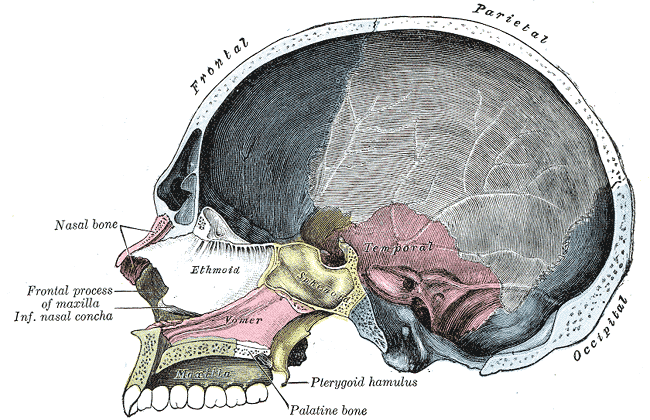
Figure 1. Saggital section of the skull. The occipital bone is at right, shown in blue.
Muscle and ligament attachments
Surface anatomy
The occipital bone, like the other cranial bones, has outer and inner tables, between which is the cancellous tissuem or diploë. The bone is especially thick at the ridges, protuberances, condyles, and anterior part of the basilar part. In the inferior fossae, it is thin, semitransparent, and destitute of diploë.
Angles
- The superior angle of the occipital bone articulates with the occipital angles of the parietal bones. In the fetal skull, this corresponds in position with the posterior fontanelle.
- The inferior angle is fused with the body of the sphenoid.
- The lateral angles are situated at the extremities of the grooves for the transverse sinuses, Each is received into the interval between the mastoid angle of the parietal and the mastoid part of the temporal.
Borders
- The superior borders extend from the superior to the lateral angles. They are deeply serrated for articulation with the occipital borders of the parietals; they form by this union the lambdoidal suture.
- The inferior borders extend from the lateral angles to the inferior angle. The upper half of each articulates with the mastoid portion of the corresponding temporal bone; the lower half with the petrous part of the same bone.
- These two portions of the inferior border are separated from one another by the jugular process, the notch on the anterior surface which forms the posterior part of the jugular foramen.
Foramen magnum
The foramen magnum is a large oval aperture with its long diameter antero-posterior. It is wider behind than in front where it is encroached upon by the condyles. The foramen magnum transmits the medulla oblongata and its membranes, the accessory nerves, the vertebral arteries, the anterior and posterior spinal arteries, and the membrana tectoria and alar ligaments.
Radiography
Physical examination
Embryology
Anomalies
Injuries/Disorders
Attachments:

Last Updated on June 27, 2024
🔗 Knowledge Graph & Clinical Correlates
OrthopaedicsOne Living Semantic Network🩺 Clinical Conditions & Indications
- Wigns There are three categories of diagnostic data collected during the physical examination (sign, symptom, and that in between)...
- Why have a privacy policy for my office Original Clinical Contribution: Authored for the OrthopaedicsOne General Orthopaedics Specialty Section. Preserved, digitized, and updated to modern evidence-based...
- Why have a practice website Original Clinical Contribution: Authored for the OrthopaedicsOne General Orthopaedics Specialty Section. Preserved, digitized, and updated to modern evidence-based...
- Who should undergo biopsy of a bone lesion Patient selection for a biopsy is important: It is intended to confirm the radiologic diagnosis in patients with...
🔬 Anatomy, Pathology & Diagnostics
- Wrist joint Wrist and Carpus
💼 Practice Management & Professional Operations
- Winthrop M. Phelps History of Orthopaedics
- Willis Cohoon Campbell History of Orthopaedics
- William Waugh History of Orthopaedics
- William Ward Plummer History of Orthopaedics
- William Thomas Gordon Pugh History of Orthopaedics
- William Stewart Halsted History of Orthopaedics
- William Stevenson Baer History of Orthopaedics
- William Morant Baker History of Orthopaedics