
1. High-Yield Overview & Pathognomonic Essence
- Radiographic Morphology: Ventral (anterior) displacement, loss of sharp demarcation, focal scalloping, or complete obliteration of the thin, radiolucent fat plane overlying the pronator quadratus muscle on true lateral wrist radiographs.
- Underlying Pathology: Highly sensitive indirect soft tissue marker of occult or non-displaced fractures of the distal radius (such as non-displaced volar rim or transverse metaphyseal fractures), distal ulna, or carpal bones (particularly scaphoid fractures), as well as acute deep volar compartment hematoma.
- Diagnostic Implication: Lowers the diagnostic threshold for cross-sectional imaging (thin-slice CT or MRI) or structured splint immobilization and scheduled radiographic re-evaluation when standard projectional radiographs fail to reveal a definitive cortical fracture line.
2. Diagnostic Sign Summary Matrix
| Primary Modality | Anatomic Location | Primary Pathology | Pathognomonic Morphology | Diagnostic Utility & Sensitivity |
|---|---|---|---|---|
| Plain Radiography (True Lateral View) | Volar distal forearm (pre-pronator fat plane) | Distal radius fractures (occult, torus, non-displaced), distal ulna fractures, carpal trauma | Ventral bowing (> 5 mm female, > 8 mm male from volar radial cortex), focal contour disruption, or complete obliteration of lucent stripe | Sensitivity ~65%, Specificity ~85%; high negative predictive value when fat stripe is crisp, linear, and undisturbed |
| Point-of-Care Ultrasound (POCUS) | Volar distal forearm acoustic window | Distal radial metaphyseal fracture, subperiosteal hematoma | Pronator Quadratus Hematoma Sign (PQHS): hypoechoic hematoma elevating muscle from cortex, with marked muscle thickening | Sensitivity ~89–94%, Specificity ~92–96% for pediatric distal forearm fractures; rapid radiation-free bedside triage |
| Computed Tomography (CT) | Distal radioulnar and radiocarpal joints | Occult cortical fractures, intra-articular micro-stepoffs, volar rim shear fractures | Hyperattenuating pronator space hematoma (40–60 HU), pronator quadratus muscle swelling, and direct visualization of cortical breach | Gold standard for fine cortical architecture; sensitivity > 98% for occult osseous disruption |
| Magnetic Resonance Imaging (MRI) | Deep volar compartment of distal forearm | Trabecular microfractures, bone marrow contusion, deep fascial disruption | T1 signal replacement of fat plane, high T2/STIR hyperintensity in pronator quadratus muscle, subperiosteal fluid, and extensive trabecular marrow edema | Sensitivity ~99–100% for radiographically occult bone marrow and soft tissue trauma |
3. Clinical Case Imaging & Standardized Figures
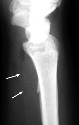
The pronator fat stripe represents a vital radiographic boundary line on the lateral wrist projection. In the non-traumatized wrist, it appears as a crisp, delicate radiolucent stripe running parallel to the volar cortex of the distal radius. Acute trauma to the distal forearm disrupts this relationship, providing an immediate soft tissue indicator of underlying skeletal injury.
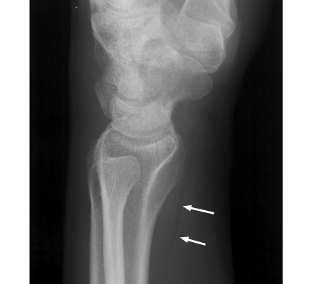
In contrast to the baseline anatomy shown in Figure 1, traumatic extravasation of blood and edema expands the deep compartment, causing obvious anterior displacement or complete erasure of the fat stripe, as demonstrated in Figure 2.
4. Pathophysiology & Biomechanical Mechanism
The anatomical basis of the pronator sign resides in the specialized osteofascial architecture of the deep volar compartment of the distal forearm. The pronator quadratus is a flat, quadrilateral muscle positioned directly against the anterior periosteum of the distal radius and ulna. It originates from the distal fourth of the anteromedial surface of the ulna and inserts broadly into the distal fourth of the anterolateral surface and volar triangular area of the radius. Innervated by the anterior interosseous nerve (AIN, a branch of the median nerve arising from C8 and T1 roots) and supplied by the anterior interosseous artery, the pronator quadratus acts as the primary dynamic pronator of the forearm and an important dynamic stabilizer of the distal radioulnar joint (DRUJ).
Covering the ventral surface of the pronator quadratus is a well-defined condensation of deep forearm fascia. Directly superficial (ventral) to this fascial sheath lies a thin layer of adipose tissue known as the pre-pronator fat plane (pronator fat stripe). This radiolucent fat cushion serves as a low-friction gliding interface separating the deep muscular belly of the pronator quadratus from the overlying long flexor tendons, specifically the flexor digitorum profundus (FDP) and flexor pollicis longus (FPL), as they converge toward the carpal canal.
The biomechanical mechanism underlying the pronator sign unfolds through the following sequence:
- Metaphyseal Fracture & Cortical Disruption: High compressive and shear loads delivered to an extended wrist (fall on an outstretched hand, FOOSH) breach the richly vascularized cancellous bone and periosteum of the distal radius or ulna.
- Pressurized Hematoma Extravasation: Disruption of metaphyseal intraosseous sinusoids and periosteal vessels extravasates arterial and venous hematoma directly into the subperiosteal space and deep pronator compartment.
- Closed Compartment Volume Expansion: Because the pronator quadratus muscle is tightly tethered to the radius and ulna by rigid osteofascial insertions, bleeding cannot freely decompress into the superficial forearm. Hydrostatic pressure within the closed pronator compartment escalates rapidly.
- Ventral Fascial Displacement: The expanding hematoma and post-traumatic muscle edema force the pronator quadratus muscle belly and its overlying deep fascial sheath ventrally. This mechanical displacement pushes the pre-pronator fat stripe anteriorly away from the volar radial cortex, producing the characteristic convex outward bowing.
- Fascial Tearing & Interstitial Infiltration: If the traumatic force tears the deep pronator fascia, or if high-energy hemorrhage infiltrates the adipose layer, the sharp radiolucent boundary is lost. This produces focal scalloping, irregular blurring, or total radiographic obliteration of the fat stripe.
5. Modality-Specific Imaging Findings
Plain Radiography
Projectional radiography remains the primary initial modality for evaluating wrist trauma. Visualization of the pronator fat stripe requires a rigorous true lateral radiograph of the wrist. The forearm must be placed in neutral rotation, with the elbow flexed to 90 degrees and the shoulder abducted to 90 degrees. Radiographic criteria for a true lateral view include exact superimposition of the distal radius and ulna, with the palmar cortex of the pisiform projecting over the central third of the scaphoid waist and scaphocapitate articulation.
On a technically adequate lateral radiograph, the normal pronator fat stripe is identifiable in approximately 90% of non-traumatized wrists. It presents as a thin, uniform radiolucent line running parallel to the distal 3 to 5 cm of the volar radial cortex, demonstrating a flat or slightly concave contour toward the bone. The normal perpendicular distance between the fat stripe and the volar cortex of the distal radius measures:
- Adult Females: Less than 5 mm.
- Adult Males: Less than 8 mm.
- Upper Limit of Normal: Distances exceeding 10 mm are universally abnormal regardless of sex or body habitus.
Pathological alterations manifest along a distinct spectrum:
- Ventral Displacement (Bowing): The fat stripe is displaced anteriorly beyond standard physiological thresholds, exhibiting a prominent convex contour directed away from the radius while maintaining a sharp margin.
- Focal Scalloping / Disruption: Localized contour defects, focal step-offs, or segmental discontinuities along the lucent line, reflecting localized hematoma breakthrough across the deep fascia.
- Complete Obliteration: Total disappearance of the radiolucent stripe, replaced by homogenous soft tissue opacity as extensive hematoma, interstitial edema, and cellular infiltration erase the fat-soft tissue density differential.
Computed Tomography
Multiplanar volumetric CT with sub-millimeter slice collimation (0.5 to 1.0 mm) provides detailed characterization of both osseous injury and soft tissue compartment changes. On axial and sagittal reconstructions, the normal pre-pronator fat plane displays characteristic negative attenuation values (-80 to -120 HU). Following trauma, acute hematoma exhibits intermediate-to-high soft tissue attenuation (40 to 60 HU), replacing the negative attenuation of the fat pad.
Cross-sectional CT readily quantifies enlargement of the pronator quadratus muscle belly compared to the uninjured contralateral forearm. More importantly, CT serves as the definitive reference modality for identifying subtle cortical infractions, non-displaced volar marginal lip fractures, occult scaphoid waist disruptions, and micro-stepoffs within the radiocarpal or distal radioulnar articular surfaces that escape detection on plain radiographs.
Magnetic Resonance Imaging & Ultrasound
Magnetic resonance imaging provides superior contrast resolution for deep soft tissues and trabecular architecture. On non-fat-suppressed T1-weighted sequences, normal pre-pronator adipose tissue produces a brilliant hyperintense stripe; extravasated blood and edema replace this signal with intermediate-to-low intensity. On fluid-sensitive sequences (T2-weighted fat-suppressed or STIR), traumatic injury manifests as intense hyperintensity within the pronator quadratus muscle, reflecting acute myoedema, intramuscular tears, or subperiosteal hematoma collections. Concurrently, STIR sequences demonstrate prominent bone marrow edema (BME) in the distal radius, distal ulna, or carpal bones, confirming occult trabecular microfractures in patients presenting with normal projectional radiographs.
High-resolution point-of-care ultrasound (POCUS), utilizing a 10 to 15 MHz linear array probe positioned longitudinally over the volar distal forearm, has established the Pronator Quadratus Hematoma Sign (PQHS). Sonographically, acute trauma produces an anechoic or hypoechoic fluid collection that elevates the deep muscle fibers off the bright specular reflector of the volar radial cortex. In pediatric emergency settings, dynamic POCUS demonstrating pronator quadratus thickening greater than 20% compared to the contralateral side achieves high diagnostic accuracy for occult buckle and greenstick fractures without exposing developing physis to ionizing radiation.
6. Diagnostic Utility, Sensitivity & Specificity
The diagnostic performance of the pronator sign has been extensively validated across contemporary systematic reviews and clinical cohort studies:
- Systematic Review & Meta-Analysis: A comprehensive meta-analysis by Kadhmawi et al. (2025) synthesizing data across 11 diagnostic cohorts (N = 2,842 patients) established a pooled sensitivity of 65% (95% CI: 58–71%) and a pooled specificity of 85% (95% CI: 79–90%). The presence of a positive pronator sign conferred a 2.44-fold increased relative risk of an underlying skeletal fracture (relative risk [RR]: 2.44, 95% CI: 1.82–3.26; positive likelihood ratio: 4.33).
- Prospective MRI Reference Standard: In a prospective blinded investigation of 74 patients with acute wrist trauma and negative or equivocal initial radiographs, Fallahi et al. (2013) demonstrated an overall sensitivity of 65% and specificity of 69%. Crucially, the authors revealed that the morphological character of the fat pad strongly influences positive predictive value: focal disruption carried a 58% fracture risk and complete obliteration carried a 54% fracture risk, whereas simple gentle raising without disruption yielded only a 38% fracture probability.
- Large-Scale Retrospective Cohort: In a landmark investigation of 1,453 consecutive wrist trauma cases, Zammit-Maempel et al. (1988) confirmed that soft tissue fat plane abnormalities accompanied the vast majority of acute distal radius fractures. The false-positive rate in uninjured control wrists was remarkably low at approximately 4%, corroborating the sign’s high diagnostic specificity.
- Technical & Rotational Sensitivity: The pronator fat stripe is exceptionally sensitive to radiographic projection geometry. A rotational deviation as small as 15 degrees in pronation or supination projects the fat plane tangentially against the radial tuberosity or pronator insertion, falsely simulating pathological displacement or causing artificial obscuration. True lateral positioning is mandatory before interpreting soft tissue displacement.
7. Differential Diagnosis & Radiographic Mimics
While an abnormal pronator fat stripe is most frequently caused by distal forearm skeletal trauma, non-osseous conditions and technical errors can produce similar radiographic appearances. Systematic differentiation is essential:
| Sign / Condition | Distinguishing Radiographic Features | Clinical Presentation | Key Differentiating Test |
|---|---|---|---|
| Acute Distal Forearm Fracture (Classic Pronator Sign) | Ventral displacement (> 5 mm female, > 8 mm male), focal disruption, or total obliteration of fat stripe; underlying cortical breach or buckle | Acute fall on outstretched hand (FOOSH), focal distal radius/ulna point tenderness, localized ecchymosis | Thin-slice multiplanar CT or high-resolution MRI revealing cortical disruption or bone marrow edema |
| Deep Forearm Space Infection (Parona Space Abscess) | Diffuse widening of deep volar soft tissues, obliteration of pronator fat plane, occasional soft tissue gas locules without cortical fracture | Spreading volar forearm pain, swelling, erythema, high fever, tenderness proximal to carpal tunnel, history of penetrating trauma or flexor tenosynovitis | Contrast-enhanced MRI showing rim-enhancing deep fluid collection; elevated inflammatory markers (ESR, CRP, leukocytosis); operative drainage and culture |
| Severe Volar Forearm Contusion / Wrist Sprain | Symmetrical, gentle widening of the pre-pronator fat plane with preserved linear margins and sharp fat-soft tissue contrast | Direct blunt impact to volar forearm or hyperextension sprain; diffuse superficial ecchymosis without focal bony point tenderness | Plain radiographs without cortical disruption; MRI demonstrating subcutaneous edema with intact cortices and absence of bone marrow edema |
| Distal Forearm Acute Compartment Syndrome | Massive, confluent soft tissue swelling of volar compartments with total obliteration of all deep and superficial intermuscular fat planes | Severe, unremitting pain out of proportion to exam, tense wooden feel of volar forearm, excruciating pain with passive finger extension, progressive sensory deficit | Intracompartmental needle manometry demonstrating delta pressure (diastolic blood pressure minus compartment pressure) < 30 mmHg; emergent fasciotomy |
| Rotational Projection Error (Technical Artifact) | Apparent widening of distance between fat plane and radius due to oblique projection; fat stripe margins remain crisp and uniform without disruption | Absence of localized trauma or mild non-specific wrist ache; normal physical examination without focal point tenderness | Repeat true lateral wrist radiograph confirming precise superimposition of distal radius and ulna and pisiform-scaphoid alignment |
8. Clinical Significance & Management Implications
The clinical value of the pronator sign lies in its role as a diagnostic red flag in emergency, primary care, and orthopaedic triage:
- Occult Fracture Triage: In a patient presenting with an acute wrist injury and focal point tenderness over the distal radius, distal ulna, or anatomical snuffbox, the presence of a positive pronator sign on otherwise normal plain radiographs should trigger presumptive fracture management. Patients should not be dismissed with an uncritical diagnosis of a minor wrist sprain. Standard clinical pathways mandate immobilization in a well-molded sugar-tong or volar splint, accompanied by early advanced imaging (thin-slice CT for cortical breaks, MRI for occult trabecular fractures and scaphoid waist injury) or planned repeat radiographic evaluation out of splint at 10 to 14 days.
- Negative Predictive Limitations: A pristine, undisturbed pronator fat stripe offers reassurance against high-energy displacement or massive compartment hematoma. However, a normal fat stripe does not completely exclude subtle undisplaced fractures, particularly pediatric torus (buckle) or incomplete greenstick fractures where the periosteal sleeve remains intact and bleeding is minimal.
- Surgical Implications & Volar Plating: During open reduction and internal fixation (ORIF) of distal radius fractures via the modified Henry approach or volar flexor carpi radialis (FCR) sheath, the pronator quadratus must be addressed. The muscle is sharply reflected from its radial insertion with an L-shaped incision, preserving a cuff of radial tissue. Following anatomical fracture reduction and plate fixation, meticulous anatomical repair of the pronator quadratus over the volar locking plate is critical. The re-approximated muscle belly creates a biological buffer that shields the overlying flexor tendons (FPL and FDP) from plate prominence and proud distal screw heads, substantially reducing the risk of postoperative flexor tenosynovitis, tendon attrition, and late FPL tendon rupture.
9. Master Class Pro-Tip
Master Class Pro-Tip: Always confirm a true lateral radiograph before diagnosing pronator sign displacement—a rotational error of just 15 degrees can simulate ventral displacement or obscure the fat stripe. If the stripe is sharply obliterated or disrupted in a symptomatic wrist without an obvious fracture line, treat as an occult fracture with immobilization and low-threshold advanced imaging (MRI or CT) rather than dismissing as a simple wrist sprain.
Master Class Radiological Pearl: Pronator Quadratus Sign Evaluation
10. Evidence-Based Synthesis
| Author & Year | Study Design | Sample Size | Key Findings & Diagnostic Accuracy | Clinical Implication | Level of Evidence | DOI |
|---|---|---|---|---|---|---|
| Kadhmawi et al. (2025) | Systematic Review & Meta-Analysis | 11 studies (N = 2,842) | Pooled sensitivity 65%, pooled specificity 85%, positive likelihood ratio 4.33; positive sign confers 2.44-fold relative risk increase for underlying fracture | Validates pronator sign as a specific clinical flag for occult wrist fractures, mandating immobilization or advanced cross-sectional imaging | Level I | DOI |
| Fallahi et al. (2013) | Prospective Blinded Diagnostic Study with MRI Reference Standard | N = 74 | Overall sensitivity 65%, specificity 69%; disrupted fat plane showed 58% fracture risk and obliterated plane showed 54% risk, whereas simple raising showed 38% | Demonstrates that qualitative morphology (disruption and obliteration) yields higher predictive value for acute fracture than simple displacement | Level II | DOI |
| Snelling et al. (2024) | Prospective Multi-Center Diagnostic Validation Study | N = 265 pediatric patients | Ultrasound pronator quadratus hematoma sign (PQHS) demonstrated sensitivity of 89.2% and specificity of 94.1% for pediatric distal forearm fractures | Establishes sonographic assessment of the pronator quadratus as a reliable bedside radiation-free diagnostic modality in pediatric trauma | Level II | DOI |
| MacEwan (1964) | Seminal Observational Cohort & Original Description | N = 400 normal & injured wrists | First described normal anatomy of the pronator fat plane and demonstrated anterior displacement or blurring in acute distal forearm fractures | Founded the radiographic sign and codified baseline anatomical parameters on lateral wrist projections | Level IV | DOI |
| Zammit-Maempel et al. (1988) | Retrospective Diagnostic Cohort | N = 1,453 | Soft tissue abnormalities accompanied the majority of distal radius fractures; false-positive rate in uninjured controls was only 4% | Confirmed high specificity of soft tissue signs in acute wrist trauma while identifying rotational artifact as a key diagnostic pitfall | Level IV | DOI |
| Zimmers (1984) | Seminal Clinical Review & Diagnostic Guide | Narrative review | Synthesized diagnostic criteria for evaluating fat plane signs in wrist and elbow trauma, standardizing emergency radiographic interpretation | Established emergency department triage protocols utilizing indirect soft tissue fat plane signs to identify occult fractures | Level V | DOI |
11. References
- MacEwan DW. Changes Due to Trauma in the Fat Plane Overlying the Pronator Quadratus Muscle: A Radiologic Sign. Radiology. 1964;82(5):879-886. DOI [Level IV: Seminal Observational Cohort]
- Zimmers TE. Fat plane radiological signs in wrist and elbow trauma. Am J Emerg Med. 1984;2(6):526-532. DOI [Level V: Expert Review]
- Zammit-Maempel I, Bisset RA, Morris J, Forbes WS. The value of soft tissue signs in wrist trauma. Clin Radiol. 1988;39(6):664-668. DOI [Level IV: Retrospective Diagnostic Cohort]
- Fallahi F, Jafari H, Jefferson G, Raby N. Explorative study of the sensitivity and specificity of the pronator quadratus fat pad sign as a predictor of subtle wrist fractures. Skeletal Radiol. 2013;42(2):249-253. DOI [Level II: Prospective Diagnostic Cohort]
- Kadhmawi I, Rashid R, Babiker M. Clinical Utility of the Pronator Quadratus Sign: A Systematic Review and Meta-Analysis. Cureus. 2025;17(12):e99766. DOI [Level I: Systematic Review & Meta-Analysis]
- Snelling PJ, Jones P, Bade D et al. Ultrasound Secondary Signs for the Diagnosis of Pediatric Distal Forearm Fractures: A Diagnostic Study. Ultrasound Med Biol. 2024;50(6):843-850. DOI [Level II: Prospective Multi-Center Diagnostic Validation Study]
Last Updated on September 28, 2026
🔗 Knowledge Graph & Clinical Correlates
OrthopaedicsOne Living Semantic Network🛠️ Operative Techniques
- Wrist Implant Arthroplasty Hand and Wrist
- Wrist disarticulation Hand and Wrist
- Wrist Dennervation Hand and Wrist
- Wrist block Pain Management
🧭 Surgical Approaches & Danger Zones
- Volar approach to the wrist Wrist and Carpus
- Volar approach to the scaphoid Wrist and Carpus
🩺 Clinical Conditions & Indications
- Wrist sprains 1. High-Yield Summary Wrist sprain is an imprecise clinical descriptor that frequently obscures high-energy ligamentous disruption of the...
- Wrist fractures Introduction Summary about significance of fracture including epidemiology. Anatomy Review the relevant anatomy of the bone, the joint,...
- Wartenberg’s syndrome 1. High-Yield Summary Wartenberg's syndrome (cheiralgia paresthetica) is an entrapment neuropathy of the superficial branch of the radial...
- Volar plate injury 1. High-Yield Summary Volar plate injuries of the proximal interphalangeal (PIP) joint represent the most frequent soft-tissue and...
🔬 Anatomy, Pathology & Diagnostics
- Wrist joint Wrist and Carpus
- Watson test Wrist and Carpus