
Contents
- Name of bone
- Location/Articulation
- Muscle and ligament attachments
- Surface anatomy
- Radiography
- Physical examination
- Embryology
- Anomalies
- Injuries/Disorders
Name of bone
Ribs
Location/Articulation
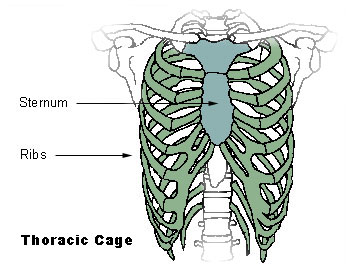
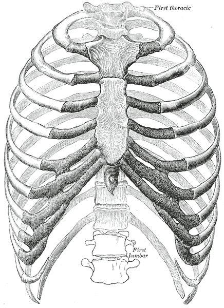
The rib cage, also known as the thoracic cage, is a bony and cartilaginous structure that surrounds the thoracic cavity and supports the pectoral girdle, forming a core portion of the human skeleton. The typical rib cage consists of 24 ribs, the sternum, costal cartilages, and the 12 thoracic vertebrae. The skin, associated fascia and muscles, and the rib cage make up the thoracic wall, providing attachments for the muscles of the neck, thorax, upper abdomen, and back.
Variations in the number of ribs occur. About 1 in 200 to 500 people have an additional cervical rib, with a female predominance. Intrathoracic supernumerary ribs are extremely rare.
Muscle and ligament attachments
Surface anatomy
All ribs are attached in the back to the thoracic vertebrae:
- The upper seven true ribs (costae verae, vertebrosternal ribs, I-VII). are attached in the front to the sternum by means of costal cartilage. Due to their elasticity, they allow movement when inhaling and exhaling.
- The 8th, 9th, and 10th ribs are called false ribs (costae spuriae, vertebrochondral ribs, VIII-X), and join with the costal cartilages of the ribs above.
- The 11th and 12th ribs are known as floating ribs (costae fluitantes, vertebral ribs, XI-XII), as they do not have any anterior connection to the sternum.

Figure 1. Rib cage
The spaces between the ribs are known as intercostal spaces; they contain the intercostal muscles, nerves, and arteries.
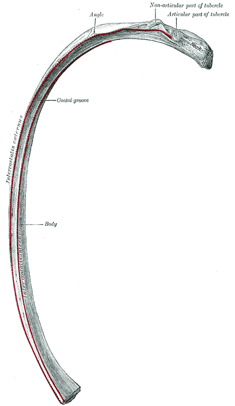
The parts of the human rib include the following:
- The head is the end of a rib closest to the vertebral column.
- The costovertebral joints are the articulations that connect the heads of the ribs to the thoracic vertebrae.
- The neck is the flattened portion that extends lateralward from the head.
- The tubercle is an eminence on the posterior surface.
- The angle is a bending part.
- The costal groove is a groove between the ridge of the internal surface of the rib and the inferior border.
Figure 2. Parts of the rib
Atypical Ribs
The atypical ribs are the 1st, 2nd, and 11th to 12th.
- The 1st rib is a shaft that is wide and nearly horizontal and has the sharpest curve of the seven true ribs. Its head has a single facet to articulate with the first thoracic vertebra (T1). It also has two grooves for the subclavian vessels, which are separated by the scalene tubercle.
- The 2nd rib is thinner, less curved, and longer than the 1st rib. It has two facets to articulate with T2 and T1, and a tubercle for muscle attachment.
- The 11th and 12th ribs have only one facet on their head. These ribs are short, with no necks or tubercles, and they terminate in the abdominal wall before fusing with the costal cartilages.
Radiography
Physical examination
Embryology
Anomalies
Abnormalities of the rib cage include pectus excavatum (“sunken chest”) and pectus carinatum (“pigeon chest”). Bifid, or bifurcated, ribs, in which the sternal end of the rib is cleaved in two, is a congenital abnormality occurring in about 1.2% of the population. The rib remnant of the 7th cervical vertebra on one or both sides is occasionally replaced by a free extra rib called a cervical rib, which can cause problems in the nerves going to the arm.
Injuries/Disorders
Rib fractures are the most common injury to the rib cage, most frequently affecting the middle ribs. Injury to several ribs can result in a flail chest.
Rib removal is the surgical excision of ribs for therapeutic or cosmetic reasons
Attachments:

Last Updated on June 27, 2024
🔗 Knowledge Graph & Clinical Correlates
OrthopaedicsOne Living Semantic Network🩺 Clinical Conditions & Indications
- Wigns There are three categories of diagnostic data collected during the physical examination (sign, symptom, and that in between)...
- Why have a privacy policy for my office Original Clinical Contribution: Authored for the OrthopaedicsOne General Orthopaedics Specialty Section. Preserved, digitized, and updated to modern evidence-based...
- Why have a practice website Original Clinical Contribution: Authored for the OrthopaedicsOne General Orthopaedics Specialty Section. Preserved, digitized, and updated to modern evidence-based...
- Who should undergo biopsy of a bone lesion Patient selection for a biopsy is important: It is intended to confirm the radiologic diagnosis in patients with...
🔬 Anatomy, Pathology & Diagnostics
- Wrist joint Wrist and Carpus
💼 Practice Management & Professional Operations
- Winthrop M. Phelps History of Orthopaedics
- Willis Cohoon Campbell History of Orthopaedics
- William Waugh History of Orthopaedics
- William Ward Plummer History of Orthopaedics
- William Thomas Gordon Pugh History of Orthopaedics
- William Stewart Halsted History of Orthopaedics
- William Stevenson Baer History of Orthopaedics
- William Morant Baker History of Orthopaedics