
1. High-Yield Overview & Pathognomonic Essence
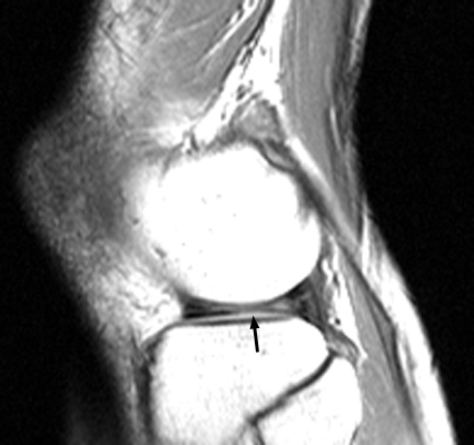
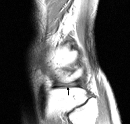
- Fundamental Radiographic Appearance: Loss of the normal contiguous bow-tie or figure-of-eight morphology of the peripheral meniscal body on sagittal magnetic resonance imaging (MRI). On standard 4 mm to 5 mm sagittal slices, an intact meniscal body spans at least two consecutive slices. The absent bow tie sign is present when zero or only one slice displays a contiguous meniscal body bridge connecting the anterior and posterior horns.
- Pathognomonic Pathology: Displaced bucket-handle meniscal tear (medial or lateral). A full-thickness longitudinal vertical cleavage tear detaches the inner central meniscal fragment (the “handle”), which displaces centrally into the intercondylar notch or anterior gutter, truncating peripheral meniscal body volume.
- Critical Diagnostic & Surgical Utility: Exhibits exceptional sensitivity (84% to 89%) and specificity (94% to 98%) for bucket-handle tears, outperforming direct fragment visualization signs such as the double PCL sign. It serves as a primary radiological indication for urgent arthroscopic reduction and meniscal repair (all-inside or inside-out) to unlock the knee, restore hoop stress distribution, and protect against accelerated chondral degradation.
2. Diagnostic Sign Summary Matrix
| Primary Modality | Anatomic Location | Primary Pathology | Pathognomonic Morphology | Diagnostic Utility & Sensitivity |
|---|---|---|---|---|
| Magnetic Resonance Imaging (MRI) | Knee / Menisci (Medial & Lateral Meniscal Body) | Displaced bucket-handle meniscal tears | Zero or only one sagittal MR slice demonstrating a contiguous meniscal body bridge connecting anterior and posterior horns (normal is ≥ 2 consecutive 4–5 mm slices) | High sensitivity (84%–89%) and high specificity (94%–98%); superior sensitivity compared to direct fragment visualization signs; dictates urgent arthroscopic reduction and repair |
3. Clinical Case Imaging & Standardized Figures

4. Pathophysiology & Biomechanical Mechanism
The normal menisci of the knee joint are specialized crescent-shaped fibrocartilaginous structures that provide critical joint congruity, shock absorption, load distribution, and joint lubrication. Microarchitecturally, the extracellular matrix consists predominantly of type I collagen fibers oriented in three distinct layers. The middle and deep zones feature dense, circumferentially oriented collagen bundles anchored to the tibial plateau by the anterior and posterior insertional meniscal root ligaments. Under axial tibiofemoral weight-bearing, compressive joint forces wedge the meniscus radially outward. The anchoring roots and intact circumferential collagen fibers resist this outward extrusion, converting vertical compressive forces into horizontal hoop stresses (tensile strain). This mechanical conversion dissipates peak articular contact forces across the femoral condyles and tibial plateaus, shielding the hyaline articular cartilage from excessive contact stresses.
A bucket-handle meniscal tear represents a longitudinally oriented, vertical full-thickness cleavage injury that propagates through the meniscal substance along the path of the circumferential fibers, extending from the posterior horn through the midbody toward the anterior horn. This tear pattern separates the meniscus into an outer peripheral rim (the “bucket”) that remains anchored to the joint capsule and tibial plateau, and a central free inner fragment (the “handle”). Because the central fragment remains attached only at its anterior and posterior extremes, repetitive rotational torque, joint fluid pressure, and tibiofemoral translation cause the fragment to buckle, flip, or translate away from the peripheral rim.
The displacement vectors of the torn fragment depend on the affected meniscus and associated ligamentous integrity. In the medial meniscus, which is larger and more tightly restrained by the deep medial collateral ligament (meniscotibial and meniscofemoral ligaments), the central fragment typically flips inward and downward into the intercondylar notch. When the anterior cruciate ligament (ACL) is intact, the displaced medial fragment comes to rest in the notch anteroinferior and parallel to the posterior cruciate ligament (PCL). In the lateral meniscus, which is smaller, more circular, and more mobile, the displaced fragment may migrate into the intercondylar notch, flip anteriorly over the anterior horn (producing a double anterior horn configuration), or displace into the popliteal hiatus.
The biomechanical basis of the absent bow tie sign stems directly from this peripheral tissue loss. Anatomically, the normal meniscal body measures 9 mm to 12 mm in transverse width in the coronal plane. When imaged on standard sagittal MRI using a slice thickness of 4 mm to 5 mm (or 3 mm slices with a 0.5 mm to 1 mm interslice gap), the peripheral meniscal body is sufficiently wide that it must appear on at least two consecutive sagittal slices as a continuous, solid band of low-signal fibrocartilage joining the anterior and posterior horns (the “bow-tie” or “figure-of-eight” appearance). When a bucket-handle tear occurs and the inner fragment displaces centrally, the remaining peripheral meniscal rim is reduced to a narrow band, typically measuring less than 4 mm to 5 mm in width. Consequently, the peripheral body can no longer span two consecutive standard sagittal slices. The resulting presence of only one slice, or zero slices, displaying meniscal body continuity constitutes the absent bow tie sign. Thus, the sign is an indirect or “subtractive” imaging sign that reliably detects meniscal volume loss regardless of where the displaced fragment has migrated.
5. Modality-Specific Imaging Findings
Plain Radiography
Standard plain radiographic evaluation of the knee includes weight-bearing anteroposterior (AP), lateral, 45-degree flexion posteroanterior weight-bearing (Rosenberg view), and axial patellofemoral (Merchant or sunrise) projections. Plain radiographs cannot directly visualize non-calcified fibrocartilage and are typically normal in the acute setting, aside from demonstrating a non-specific joint effusion on the lateral view (suprapatellar pouch distension > 5 mm). However, plain radiography plays an indispensable triage role:
- Exclusion of Osseous Pathology: Radiographs rule out tibial spine avulsion fractures, osteochondral fractures with intra-articular loose bodies, patellar dislocation remnants, and acute plateau fractures that can clinically present as acute mechanical joint locking.
- Associated Avulsion Fractures: A lateral capsular avulsion fracture (Segond fracture) indicates high-grade anterolateral complex and anterior cruciate ligament injury, which is associated with bucket-handle tears in 30% to 50% of acute traumatic presentations.
- Chronic Degenerative Sequelae: In chronic or neglected bucket-handle tears, the loss of hoop strain dissipation produces progressive joint space narrowing, subchondral sclerosis, and marginal osteophytosis localized to the affected compartment on the 45-degree flexion Rosenberg view. Significant pre-existing osteoarthritis (Kellgren-Lawrence grade 3 or 4) substantially reduces the prognosis of meniscal repair and shifts the surgical algorithm toward meniscectomy, realignment osteotomy, or arthroplasty.
Computed Tomography
Non-contrast computed tomography (CT) has poor intrinsic soft-tissue contrast resolution and cannot reliably differentiate the meniscal fibrocartilage from surrounding synovial fluid and capsule. Consequently, non-contrast CT is not recommended for primary meniscal evaluation. However, CT arthrography (direct intra-articular injection of iodinated contrast under fluoroscopic or sonographic guidance) serves as an effective diagnostic alternative for patients with absolute contraindications to MRI (e.g., non-MRI-compatible cardiac pacemakers, neurostimulators, or metallic orbital foreign bodies):
- Sub-Millimeter Multiplanar Reconstructions: Thin-slice volumetric CT arthrography (0.5 mm to 0.75 mm slice thickness) provides exceptional spatial resolution, delineating the contrast medium dissecting into the longitudinal cleavage plane between the peripheral rim and the displaced fragment.
- Morphologic Mapping: Multiplanar coronal and sagittal reformations clearly demonstrate the absence of the normal peripheral meniscal body width and locate the hyperattenuating contrast-outlined displaced fragment within the intercondylar notch or anterior recess. CT arthrography is also highly accurate in assessing adjacent hyaline articular cartilage thickness and focal chondral delamination.
Magnetic Resonance Imaging & Ultrasound
Magnetic resonance imaging (MRI) is the definitive diagnostic gold standard for meniscal pathology. Examination protocols must include high-resolution proton density (PD) weighted and T2-weighted fast spin-echo (FSE) sequences in sagittal, coronal, and axial planes, ideally with fat suppression. Sagittal images should be acquired with a slice thickness of 4 mm to 5 mm (or 3 mm slices with minimal spacing) to maintain reliable bow-tie slice counting.
On sagittal MR images, normal meniscal tissue displays uniform low signal intensity (signal void) on all pulse sequences. When scrolling from the periphery toward the center of the joint, the outer 9 mm to 12 mm of the meniscal body appears as a solid bow-tie connecting the anterior and posterior triangular horns. Meeting the diagnostic criterion for the absent bow tie sign requires that zero or only one sagittal slice demonstrates this contiguous bow-tie configuration. In addition to the absent bow tie sign, MRI must be systematically scrutinized for associated direct and indirect signs of displaced bucket-handle tears:
- Double PCL Sign: Visualized on sagittal views when a displaced medial meniscal fragment lies parallel and anteroinferior to the posterior cruciate ligament within the intercondylar notch. This sign is highly specific (98% to 100%) but occurs only when the ACL is intact and the fragment is medial and sufficiently long.
- Double Anterior Horn Sign: Visualized on sagittal views when the displaced fragment flips anteriorly to lie on top of or adjacent to the anterior horn, creating the appearance of two anterior horns (one anatomic and one displaced fragment). This is particularly common in lateral meniscal tears.
- Fragment in the Notch Sign: Best visualized on coronal and axial PD-weighted images as a distinct, low-signal band of fibrocartilaginous tissue residing within the intercondylar notch, separate from the anterior and posterior cruciate ligaments.
- Truncated Triangle / Flipped Fragment Sign: On coronal images, the peripheral meniscal rim appears abnormally truncated, blunted, or diminutive (measuring < 5 mm in width), accompanied by visualization of the displaced fragment displaced into the intercondylar space.
- Disproportionate Horn Sign: On sagittal images, a marked discrepancy in the relative size of the anterior versus posterior horn (e.g., an abnormally diminutive posterior horn with a disproportionately enlarged anterior horn) indicates that a segment has detached and flipped.
Diagnostic musculoskeletal ultrasound (US) utilizing high-frequency linear-array transducers (12 MHz to 18 MHz) can evaluate the peripheral meniscus at the joint line. Dynamic sonography may identify joint effusion, parameniscal cysts, meniscal extrusion (> 3 mm beyond the tibial margin), and an abrupt loss or blunting of the normal triangular hyperechoic peripheral meniscal profile. However, ultrasound cannot penetrate the osseous acoustic barrier of the femoral condyles and intercondylar notch, making it incapable of visualizing centrally displaced fragments or reliably assessing the absent bow tie sign. Ultrasound is therefore reserved for bedside screening of superficial ligamentous pathology and effusion aspiration, rather than definitive meniscal staging.
6. Diagnostic Utility, Sensitivity & Specificity
The absent bow tie sign is widely recognized as the single most sensitive magnetic resonance imaging sign for the detection of bucket-handle meniscal tears. In the landmark original validation study by Helms, Laorr, and Cannon (1998), the absent bow tie sign demonstrated a sensitivity of 88% and a specificity of 98% across 33 bucket-handle tears and 115 intact controls. Subsequent independent clinical investigations by Watt et al. (2000) confirmed a sensitivity of 84% and specificity of 94% in a cohort of 75 knees. Dorsay and Helms (2003) comparatively evaluated seven distinct MR signs in 138 patients, establishing that the absent bow tie sign achieved the highest overall diagnostic sensitivity (88.7%) and specificity (97.8%) among all evaluated parameters. When the absent bow tie sign was combined with direct visualization of a fragment in the notch, the positive predictive value (PPV) reached 98%.
The superior sensitivity of the absent bow tie sign becomes clinically apparent when contrasted with direct fragment visualization signs. In the retrospective correlation study by Ververidis et al. (2006) involving 65 arthroscopically confirmed bucket-handle tears, the absent bow tie sign demonstrated 89.2% sensitivity, significantly outperforming the double PCL sign (44.6%), the double anterior horn sign (33.8%), and the flipped meniscus sign (27.7%). While signs such as the double PCL sign exhibit exceptional specificity approaching 100%, their clinical sensitivity is severely limited by strict anatomical prerequisites: the tear must involve the medial meniscus, the torn fragment must be sufficiently large to traverse the notch, the anterior cruciate ligament must remain intact to prevent abnormal notch kinematics, and the fragment must displace inferiorly rather than anteriorly or into a gutter. In sharp contrast, because the absent bow tie sign is an indirect “subtractive” sign assessing peripheral tissue absence rather than fragment localization, it remains positive regardless of whether the torn fragment is medial, lateral, intact-ACL, ACL-deficient, flipped anteriorly, or fragmented within the notch.
Technical parameters strongly influence the interpretation of the absent bow tie sign. The classic diagnostic threshold of at least two contiguous bow-tie slices is strictly calibrated for standard sagittal slice thicknesses of 4.0 mm to 5.0 mm. When modern thin-slice protocols (2.0 mm to 3.0 mm) or 3D isotropic volumetric acquisitions are utilized, a normal meniscal body may span three, four, or five consecutive slices; under such acquisition parameters, an absolute slice count of two is abnormal, and radiologists must normalize the slice count to the millimeter width of the peripheral body (normal width 9 mm to 12 mm). Furthermore, false-positive interpretations can occur in small stature adults, pediatric patients, or following previous partial meniscectomy, necessitating careful clinical history correlation and anatomic measurement.
7. Differential Diagnosis & Radiographic Mimics
| Sign / Condition | Distinguishing Radiographic Features | Clinical Presentation | Key Differentiating Test |
|---|---|---|---|
| Classic Absent Bow Tie Sign (Bucket-Handle Tear) | Zero or only 1 contiguous sagittal bow-tie slice on 4–5 mm MRI; displaced low-signal fragment identified in intercondylar notch or anterior gutter; truncated peripheral rim | Acute twisting injury, sudden joint effusion, springy mechanical block to terminal extension (locked knee), joint line tenderness | Proton density / T2-weighted MRI (sagittal and coronal sequences); diagnostic knee arthroscopy confirming bucket-handle detachment |
| Prior Partial Meniscectomy | Single or absent bow-tie slice on sagittal MRI; smooth, well-healed, truncated meniscal body contour without an adjacent vertical tear; zero displaced fragments in the intercondylar notch or gutters | History of prior knee arthroscopy; surgical portal scars visible on physical exam; recurrent joint pain or joint line tenderness without acute mechanical locking | Detailed surgical history; operative report review; absence of displaced fragment or jagged cleavage planes on multiplanar MRI |
| Small / Pediatric Knee (Slice Thickness Volume Artifact) | Single bow-tie slice visualized on standard 4–5 mm sagittal MRI due to small absolute meniscal body width (< 9 mm); completely normal triangular morphology of anterior and posterior horns; zero displaced fragment in notch | Pediatric patient or adult of diminutive stature; no acute high-energy trauma; full range of motion without joint locking or mechanical catch | Thin-slice sagittal MRI protocol (≤ 2.0–3.0 mm slices) or 3D isotropic acquisition demonstrating multiple contiguous bow-tie slices across the narrow body |
| Complete Radial Tear of Meniscal Body | Focal transverse cleft running perpendicular to the circumferential fibers; produces a “ghost meniscus” or empty space on one sagittal slice; anterior and posterior horn volumes remain normal without a displaced fragment in the notch | Acute rotational pop or chronic degenerative loading; localized joint line pain; joint effusion; typically lacks the springy mechanical block to extension | Coronal and axial MRI displaying vertical radial cleavage through the body width with normal horn volume and zero fragment displacement |
| Discoid Meniscus with Secondary Tear | Abnormally widened meniscal body demonstrating ≥ 3 contiguous bow-tie slices on standard 4–5 mm sagittal cuts (pre-tear); central tear may produce abnormal central fragments, but peripheral body remains abnormally hypertrophic | Adolescent or young adult; chronic lateral joint clicking, popping, or snapping knee syndrome; lateral joint line tenderness; variable range of motion loss | Coronal MRI demonstrating meniscal tissue extending continuously into the intercondylar notch across multiple slices (> 14 mm coronal width) |
| Congenital Meniscal Hypoplasia / Aplasia | Extremely rare congenital underdevelopment or absence of the meniscal body (most commonly lateral); diminutive bow tie without an associated tear, displaced fragment, or joint effusion | Asymptomatic incidental finding or mild mechanical instability; childhood presentation; absence of traumatic injury history | Bilateral comparative knee MRI or high-resolution 3D imaging demonstrating symmetric or isolated developmental hypoplasia with intact capsular attachments |
8. Clinical Significance & Management Implications
The absent bow tie sign is a critical radiological marker of a displaced bucket-handle meniscal tear, carrying major surgical and prognostic ramifications that directly dictate clinical management:
- The Acute “Locked Knee” Emergency: When a bucket-handle fragment displaces into the intercondylar notch, it physically blocks full femoral condylar roll-and-glide kinematics, producing an acute “locked knee” characterized by a springy block to terminal passive extension. Persistent mechanical locking subjects the articular cartilage of the femoral condyle and tibial plateau to localized shear stress, leading to rapid chondral indentation, scuffing, and accelerated chondrolysis. Furthermore, prolonged fixed flexion contracture produces secondary hamstring spasm and capsular contracture, complicating subsequent surgical reduction.
- Closed Reduction Maneuvers: In the acute emergency department or office setting, gentle closed manipulation may be attempted under intra-articular local anesthesia or conscious sedation. The clinician applies axial traction with the knee in 90 degrees of flexion, alternates internal and external tibial rotation, and gently brings the joint into passive extension. Successful reduction is heralded by an audible or palpable clunk and immediate restoration of full, pain-free extension. However, closed reduction does not heal the underlying vertical tear; spontaneous re-displacement is extraordinarily high, and surgical stabilization remains definitively indicated.
- Urgent Operative Timing & Prevention of Plastic Deformation: A displaced bucket-handle tear identified by an absent bow tie sign represents an urgent surgical indication. Multiple clinical trials demonstrate that surgical intervention delayed beyond 6 to 12 weeks results in progressive plastic deformation, retraction, fraying, and fibrosis of the displaced handle. Chronically deformed fragments become rigid and shortened, rendering anatomical reduction back into the peripheral vascular bed impossible and forcing the surgeon to perform a subtotal meniscectomy. Early surgery (ideally within 1 to 3 weeks of injury) optimizes tissue viability and repairability.
- Arthroscopic Repair Modalities (All-Inside, Inside-Out, and Outside-In Techniques): Modern orthopaedic practice prioritizes meniscal preservation over resection whenever tissue quality allows. Fourth-generation low-profile all-inside suture anchors with self-sliding knots are ideal for securing tears of the posterior horn and posterior third of the meniscal body, avoiding posterior neurovascular risks. Inside-out suture repair remains the gold standard for repairs of the midbody and junction of the middle and anterior thirds, utilizing flexible double-loaded cannulated needles passed from inside the joint out through the capsule and tied over dedicated mini-open safety incisions (posteromedial incision to protect the saphenous neurovascular bundle; posterolateral incision to protect the common peroneal nerve). Outside-in suture repair is optimal for securing far anterior horn tears under direct visualization.
- Biologic Augmentation of the Meniscal Repair Bed: Given that many bucket-handle tears propagate near or across the red-white junction, biologic stimulation is critical to promote vascular healing. Standard adjuncts include mechanical trephination of the peripheral rim, synovial rasping, intercondylar notch microfracture (marrow venting) to release mesenchymal stem cells and growth factors, and application of autologous platelet-rich plasma (PRP) or fibrin clot. Concomitant ACL reconstruction significantly enhances meniscal healing rates (healing rates exceed 85% to 90% when performed concurrently with ACL reconstruction, compared to 60% to 70% in isolated repairs).
- Long-Term Chondroprotection vs. Partial Meniscectomy: Resection of a bucket-handle fragment removes up to 50% to 70% of the meniscal volume. Biomechanical investigations demonstrate that subtotal meniscectomy completely abolishes hoop strain transmission, reducing tibiofemoral contact area by 50% to 70% and increasing peak local contact stresses by 200% to 300%. As synthesized in the systematic review by Ross et al. (2025), patients undergoing meniscal repair exhibit substantially lower rates of radiographic osteoarthritis, superior patient-reported outcome measures (IKDC, KOOS), and higher rates of return to pre-injury sports at 10 to 15 years compared to patients managed with partial meniscectomy. Partial meniscectomy is strictly reserved for severely macerated, degenerate, or chronically deformed fragments that cannot be anatomically reduced into the peripheral bed.
9. Master Class Pro-Tip
Master Class Pro-Tip: When reviewing sagittal knee MR images for suspected meniscal tears, always count the meniscal body slices from the peripheral joint capsule inward before searching for a displaced fragment in the notch. If you identify only zero or one bow-tie slice on standard 4 mm to 5 mm sagittal cuts, an absent bow tie sign is established—meaning tissue is definitively missing from the peripheral joint margin. Never abandon your suspicion of a bucket-handle tear simply because the classic double PCL sign is absent; in lateral meniscal tears, or in an ACL-deficient knee where abnormal tibiofemoral translation alters notch kinematics, the displaced fragment rarely positions itself neatly beneath the PCL. Instead, systematically scrutinize the anterior compartment for a double anterior horn sign, inspect the intercondylar notch on coronal and axial views for an amorphous low-signal fragment, and examine the popliteal hiatus for flipped meniscal tissue.
Master Class Radiological Pearl: Systematic Meniscal Body Slice Counting
10. Evidence-Based Synthesis
| Author & Year | Study Design | Sample Size | Key Findings & Diagnostic Accuracy | Clinical Implication | Level of Evidence | DOI |
|---|---|---|---|---|---|---|
| Helms CA, Laorr A, Cannon WD Jr. (1998) | Retrospective Cohort Study | N = 33 bucket-handle tears (115 normal controls) | Original description of the absent bow tie sign (defined as fewer than two contiguous sagittal slices showing normal meniscal body). Demonstrated 88% sensitivity and 98% specificity for bucket-handle tears. | Establishes loss of peripheral meniscal body volume as a primary diagnostic indicator of fragment displacement into the notch. | Level IV | DOI |
| Watt AJ, Halliday T, Raby N. (2000) | Retrospective Comparative Validation Study | N = 75 knees (25 bucket-handle tears, 50 non-displaced/intact controls) | Confirmed high diagnostic utility of the absent bow tie sign with 84% sensitivity and 94% specificity. Noted superior sensitivity compared to the double PCL sign. | Demonstrates that peripheral body volume loss frequently precedes recognition of centrally displaced fragments. | Level III | DOI |
| Dorsay TA, Helms CA. (2003) | Retrospective Comparative Diagnostic Study | N = 138 patients (46 bucket-handle tears, 92 controls) | Comparative appraisal of 7 MRI signs. Absent bow tie sign demonstrated the highest sensitivity (88.7%) among all signs, with 97.8% specificity. Combination of absent bow tie and fragment in notch yielded 98% PPV. | Validates the absent bow tie sign as the most sensitive single MR sign for meniscal bucket-handle displacement. | Level III | DOI |
| Aydingöz U, Firat AK, Atay OA, et al. (2003) | Diagnostic Cohort Study & Systematic Review | N = 42 arthroscopically proven bucket-handle tears | Correlated MRI signs with tear displacement patterns. Absent bow tie sign was present in 88% of cases, correlating with fragment migration into intercondylar notch or anterior horn. | Guides preoperative arthroscopic portal selection and repair planning based on displacement vector. | Level IV | DOI |
| Ververidis AN, Verettas DA, Kazakos KJ, et al. (2006) | Retrospective Correlation Study | N = 65 patients with arthroscopically confirmed tears | Evaluated 6 MRI signs against arthroscopy. Absent bow tie sign exhibited 89.2% sensitivity, outperforming double PCL (44.6%) and double anterior horn (33.8%) signs. | Emphasizes that absence of normal peripheral meniscal tissue is more dependable than detecting the displaced fragment itself. | Level IV | DOI |
| Ross P, Livingston M, Saraf SM, et al. (2025) | Systematic Review & Meta-Analysis | Systematic review of 48 studies (N = 2,410 patients) | Comprehensive synthesis of modern diagnostic criteria and repair outcomes. MRI sensitivity for bucket-handle tears pooled at 86-90% when absent bow tie sign is present. Meniscal preservation via repair yielded superior long-term chondroprotection compared to partial meniscectomy. | Confirms urgent reduction and repair indications for displaced bucket-handle fragments to prevent permanent chondral breakdown. | Level II | DOI |
11. References
- Helms CA, Laorr A, Cannon WD Jr. The absent bow tie sign in bucket-handle tears of the menisci in the knee. AJR Am J Roentgenol. 1998;170(1):57-61. DOI [Level IV: Retrospective Cohort Study]
- Watt AJ, Halliday T, Raby N. The value of the absent bow tie sign in MRI of bucket-handle tears. Clin Radiol. 2000;55(8):622-626. DOI [Level III: Retrospective Comparative Validation Study]
- Dorsay TA, Helms CA. Bucket-handle meniscal tears of the knee: sensitivity and specificity of MRI signs. Skeletal Radiol. 2003;32(5):266-272. DOI [Level III: Retrospective Comparative Diagnostic Study]
- Aydingöz U, Firat AK, Atay OA, et al. MR imaging of meniscal bucket-handle tears: a review of signs and their relation to arthroscopic classification. Eur Radiol. 2003;13(3):618-625. DOI [Level IV: Diagnostic Cohort Study & Systematic Review]
- Ververidis AN, Verettas DA, Kazakos KJ, et al. Meniscal bucket handle tears: a retrospective study of arthroscopy and the relation to MRI. Knee Surg Sports Traumatol Arthrosc. 2006;14(4):343-349. DOI [Level IV: Retrospective Correlation Study]
- Ross P, Livingston M, Saraf SM, et al. Bucket-Handle Meniscus Tears: Epidemiology, Diagnosis, Management, and Outcomes. JBJS Rev. 2025;13(12):e25.00144. DOI [Level II: Systematic Review & Meta-Analysis]
Last Updated on September 29, 2026
🔗 Knowledge Graph & Clinical Correlates
OrthopaedicsOne Living Semantic Network🛠️ Operative Techniques
- Valgus osteotomy for development coxa vara Hip and Knee
- Unicondylar Knee Arthroplasty Joint Reconstruction
- Unicompartmental Knee Arthroplasty – An Option for Treating Unicompartmental Osteoarthritis Joint Reconstruction
- Triple innominate osteotomy Hip and Knee
🩺 Clinical Conditions & Indications
- Valgus-Unloader Bracing in Knee Osteoarthritis Original Clinical Contribution: Authored for the OrthopaedicsOne Adult Reconstruction Specialty Section. Preserved, digitized, and updated to modern evidence-based...
- Valgus deformity of the knee Valgus deformity is a term for outward angulation of the distal segment of a bone or joint. The...
🔬 Anatomy, Pathology & Diagnostics
- What Is the Latest Update on Hip Impingement Hip Joint & Proximal Femur
- What Is the Latest Update on Hip Dysplasia Hip Joint & Proximal Femur
- What Is the Evidence-Based Data for Gender Specific Arthroplasty in TKA Knee Joint & Extensor Mechanism
- Warfarin – What Is Our Goal Knee Joint & Extensor Mechanism
💼 Practice Management & Professional Operations
- What Is the Evidence-Based Data for Mobile Bearing Knees Mobile and Wireless