
1. High-Yield Overview & Pathognomonic Essence
- Morphological Hallmark: The “Anteater Nose” sign represents a prominent, tubular, or spatulate elongation of the anterior process of the calcaneus (calcaneal rostrum) extending anterosuperiorly toward the lateral aspect of the navicular bone on lateral weight-bearing radiographs of the foot.
- Pathognomonic Significance: This projection serves as the classic direct radiographic indicator of calcaneonavicular coalition (whether fibrous syndesmosis, cartilaginous synchondrosis, or complete osseous synostosis), representing an abnormal developmental bridging across the midtarsal interval.
- Critical Diagnostic & Surgical Utility: Identification of the sign provides rapid confirmation of the underlying etiology in pediatric and adolescent patients presenting with rigid pes planovalgus (“peroneal spastic flatfoot”) and recurrent ankle inversion injuries, triggering targeted cross-sectional imaging (CT or MRI) to delineate coalition composition, quantify bar surface area, and plan surgical resection with interposition.
2. Diagnostic Sign Summary Matrix
| Primary Modality | Anatomic Location | Primary Pathology | Pathognomonic Morphology | Diagnostic Utility & Sensitivity |
|---|---|---|---|---|
| Plain Radiography (Lateral Weight-Bearing Foot) | Calcaneonavicular interval (anterior calcaneal process to posterolateral navicular body) | Calcaneonavicular Coalition (Synostosis, Synchondrosis, or Syndesmosis) | Elongated, tubular anterior calcaneal process projecting anterosuperiorly toward navicular (“Anteater Nose”) | High direct screening sensitivity (80-86%) and specificity (90%); primary first-line radiographic screening view |
| Plain Radiography (45-Degree Medial Oblique / Slomann View) | Calcaneonavicular joint interface | Calcaneonavicular Coalition | Articular joint space narrowing, irregular sclerotic margins, or solid osseous continuity across the bar | Superior sensitivity (92%) on plain radiography; directly profiles the pseudarthrosis or bony synostosis |
| Computed Tomography (CT) | Calcaneocuboid-navicular transition and anterior subtalar complex | Calcaneonavicular Coalition (Osseous or Fibrocartilaginous) | Continuous osseous bridge (synostosis) or narrow, irregular joint space with subchondral sclerosis and microcysts | Gold standard for osseous architecture (sensitivity >95-98%, specificity 98-100%); maps bar surface area (<50% vs >50%) |
| Magnetic Resonance Imaging (MRI) | Extracapsular calcaneonavicular syndesmosis or synchondrosis | Fibrous or Cartilaginous Tarsal Coalition | Low-to-intermediate signal bar bridging bones, marked adjacent subchondral bone marrow edema on T2 FS / STIR | Unmatched sensitivity (95-100%) for non-osseous coalitions in pre-ossified pediatric cohorts and stress marrow edema |
3. Clinical Case Imaging & Standardized Figures
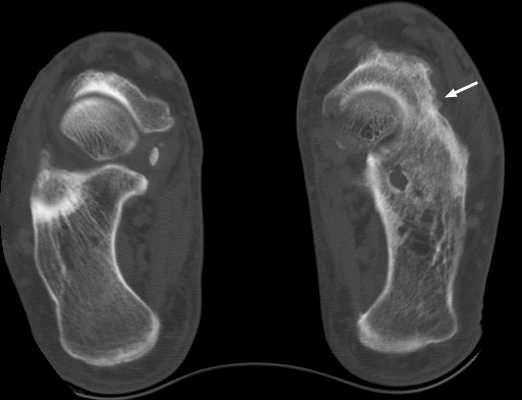
Cross-sectional imaging provides definitive structural assessment of the tarsal architecture. In Figure 1, reformatted computed tomography clearly delineates the continuous osseous integration and pseudarthrotic articulation between the calcaneus and navicular, demonstrating adjacent subchondral sclerosis and cortical irregularity that distinguish true coalition from simple anatomical elongation.
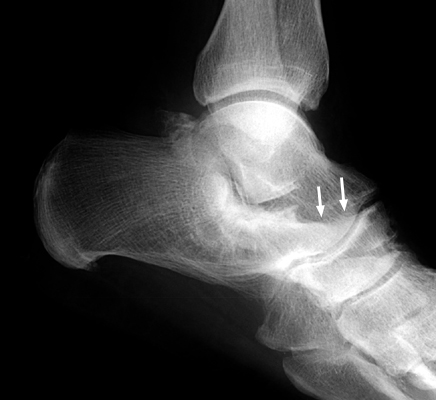
Weight-bearing lateral plain radiography serves as the primary screening examination for tarsal coalition. In Figure 2, the anterior calcaneal process demonstrates abnormal anterosuperior elongation, extending past the anterior margin of the cuboid to approach the lateral aspect of the navicular, forming the classical tubular silhouette resembling an anteater’s snout.
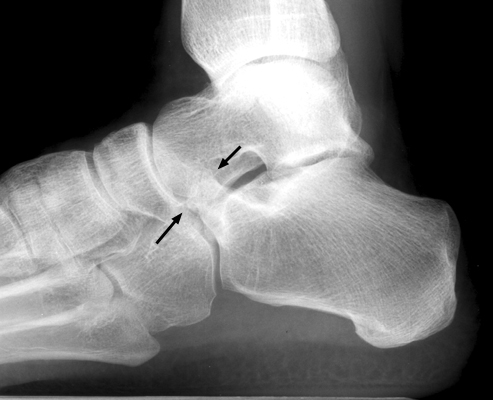
The 45-degree medial oblique (internal oblique / Slomann) projection profiles the calcaneonavicular space tangential to the beam. In Figure 3, the abnormal bar and its narrowed, sclerotic pseudarthrosis are brought into sharp relief, revealing the loss of normal separation between the calcaneal beak and the navicular.
4. Pathophysiology & Biomechanical Mechanism
Tarsal coalitions represent congenital developmental anomalies originating from the failure of normal mesenchymal differentiation and segmentation in the embryonic tarsal blastema during the 4th to 8th weeks of gestation. In normal embryogenesis, the contiguous cartilaginous tarsal anlage undergoes selective apoptosis and cavitation to produce distinct synovial joint cavities. When this programmed cleavage fails, a persistent mesenchymal bridge remains between adjacent tarsal elements, most commonly involving the calcaneonavicular interval or the middle facet of the talocalcaneal joint.
During infancy and early childhood, this congenital bridge consists of flexible, fibroblastic syndesmotic tissue or a cartilaginous synchondrosis. Because cartilage and fibrous tissue possess low elastic moduli and accommodate mechanical deformation under load, young children remain asymptomatic, retaining supple hindfoot kinematics. However, between 8 and 12 years of age (and up to 14 years), the coalition undergoes progressive endochondral ossification, synchronizing with the physiological ossification timetable of the anterior process of the calcaneus. As the coalition progressively stiffens and converts into rigid bone (synostosis) or an unyielding synchondrosis, foot mechanics alter drastically.
The biomechanical hallmark of calcaneonavicular coalition is rigid restriction or complete abolition of normal subtalar joint motion. Under physiological gait conditions, the subtalar joint executes 20 to 30 degrees of inversion and approximately 10 degrees of eversion. During initial contact and loading response, subtalar eversion allows midtarsal joint axes (talonavicular and calcaneocuboid) to parallelize, making the foot flexible for shock absorption. In terminal stance, subtalar inversion diverts these axes into divergence, converting the midfoot into a rigid lever arm for efficient forward propulsion.
When the calcaneonavicular bar bridges the hindfoot to the midfoot, it locks the calcaneus to the navicular, eliminating subtalar inversion and eversion. In the absence of normal triplanar motion, the foot adopts a compensatory fixed valgus posture. Ground reaction forces induce chronic collapse of the medial longitudinal arch, placing severe traction on the plantar calcaneonavicular (spring) ligament, long plantar ligament, and posterior tibial tendon.
Simultaneously, rigid hindfoot limitation and lateral impingement provoke reflexive muscular splinting. The peroneus brevis and longus muscles contract persistently to guard against painful subtalar rotational stresses. This involuntary protective spasm, termed “peroneal spastic flatfoot,” eventually matures into adaptive, structural myotendinous shortening and contracture, locking the hindfoot into permanent valgus. The resulting mechanical rigidity shifts pathological shear forces across the transverse tarsal (Chopart) joints and posterior subtalar facet, producing dorsal talonavicular traction spurring, subchondral microfractures, intense bone marrow edema, and accelerated secondary degenerative arthrosis.
5. Modality-Specific Imaging Findings
Plain Radiography
The standard radiographic evaluation of suspected tarsal coalition comprises weight-bearing anteroposterior (AP), lateral, and 45-degree medial oblique (internal oblique / Slomann) views of the foot.
- Lateral Weight-Bearing Radiograph: The classic “Anteater Nose” sign manifests as a prominent, tubular, or spatulate elongation of the anterior process of the calcaneus extending anterosuperiorly toward the lateral aspect of the navicular. The elongated rostrum projects past the anterior articular facet of the cuboid, mimicking the snout of an anteater (Myrmecophaga tridactyla). In fibrous or cartilaginous coalitions, the cortices of the opposing bones are irregular and sclerotic, separated by a narrowed pseudarthrosis. In complete synostosis, continuous trabecular architecture traverses the calcaneonavicular interval.
- 45-Degree Medial Oblique (Slomann) View: This projection represents the single most accurate plain radiographic view for evaluating calcaneonavicular coalition. By directing the X-ray beam parallel to the plane of the calcaneonavicular articulation, the bar is unmasked without anatomical overlap. In bony synostosis, solid osseous bridging is directly visualized. In fibrous or cartilaginous coalitions, the articulation demonstrates marked joint space narrowing (typically <2 mm), subchondral sclerosis, and irregular, wavy cortical interdigitation.
- Anteroposterior (AP) View: While the coalition itself is obscured by overlapping tarsal bones on the AP view, this projection provides critical assessment of secondary peritalar subluxation, revealing forefoot abduction, midtarsal breach, and lateral uncoverage of the talar head exceeding the normal threshold of 30%.
- Secondary Radiographic Signs:
• Dorsal Talar Beaking: A traction spur arising at the dorsal aspect of the talonavicular joint margin, provoked by abnormal elevation and sagittal subluxation of the navicular over the talar head during attempted gait motion. This must be distinguished from true degenerative osteophytes, as it initially reflects traction rather than loss of talonavicular articular cartilage.
• Narrowing of the Posterior Subtalar Facet: Results from chronic biomechanical stress concentration and restricted subtalar glide.
• Asymmetry of the Anterior Subtalar Facet: Hypoplasia or aberrant positioning of the anterior subtalar articulation.
• Disruption of the Cyma Line: Loss of the normal smooth, unbroken S-shaped curve formed by the talonavicular and calcaneocuboid joint lines on AP and lateral projections.
Computed Tomography
Computed tomography (CT) serves as the definitive structural gold standard for characterizing tarsal coalitions and planning surgical intervention. Thin-slice volumetric CT acquisitions (≤1.0 mm slice thickness) with multiplanar 2D reformations (sagittal, coronal, axial) and 3D volume renderings provide complete spatial characterization of the coalition.
- Morphological Delineation: Sagittal reformations demonstrate the longitudinal reach and anterosuperior trajectory of the anterior calcaneal process toward the navicular, while axial and oblique reformations document the mediolateral width and thickness of the bridging bar. In complete osseous synostosis, CT displays confluent, mature cortical and trabecular continuity crossing the calcaneonavicular space. In fibrous or cartilaginous coalitions (syndesmosis or synchondrosis), CT demonstrates joint space narrowing, marked subchondral sclerosis, cortical irregularity, and subchondral microcystic changes.
- Preoperative Quantitative Bar Mapping: High-resolution CT allows accurate quantitative measurement of the cross-sectional surface area of the coalition relative to the posterior subtalar and calcaneocuboid articulations. Coalition size is a primary surgical prognostic indicator: coalitions involving <50% of the joint surface are amenable to bar resection, whereas coalitions exceeding 50% carry high failure rates after resection and warrant primary arthrodesis.
- Assessment of Adjacent Degenerative Arthritis: CT definitively detects subtle subchondral sclerosis, cyst formation, and joint space loss at the posterior subtalar, talonavicular, and calcaneocuboid joints, ruling out secondary arthrosis that would contraindicate isolated bar excision.
Magnetic Resonance Imaging & Ultrasound
Magnetic resonance imaging (MRI) is the premier modality for identifying non-osseous coalitions in young children before complete ossification occurs and for detecting acute mechanical stress responses in symptomatic patients.
- Signal Characteristics of Coalition Tissue: On T1-weighted sequences, fibrous tissue presents as uniform low signal intensity, while cartilaginous tissue demonstrates intermediate signal intensity continuous between the anterior calcaneal process and the lateral navicular margin. In mature osseous synostosis, T1 sequences show continuous fatty bone marrow signal traversing the bridge uninterrupted. Fluid-sensitive sequences (T2-weighted fat-suppressed, PD fat-suppressed, and STIR) demonstrate intermediate-to-high signal in cartilaginous synchondroses and low signal in fibrous syndesmoses.
- Subchondral Bone Marrow Edema: A paramount diagnostic feature on fluid-sensitive MRI sequences is extensive, high-signal bone marrow edema within the anterior calcaneal process and the adjacent posterolateral navicular body. This edema reflects active microtrabecular bone fatigue, abnormal shear stress, and repetitive mechanical impaction, correlating strongly with clinical pain and symptom severity.
- Soft Tissue and Chondral Evaluation: MRI reliably evaluates adjacent soft tissue structures, including peroneal tenosynovitis or subluxation, posterior tibial tendinopathy, and tears of the cervical or interosseous talocalcaneal ligaments within the sinus tarsi. Chondral maps confirm the preservation or thinning of articular cartilage in adjacent hindfoot joints.
- High-Resolution Ultrasound: Dynamic musculoskeletal sonography identifies non-osseous bridging as an irregular, hypoechoic soft tissue band bridging the interruption between the hyperechoic cortical margins of the calcaneus and navicular. Direct transducer pressure over the sonographically identified bar elicits localized pain, confirming the symptomatic locus.
6. Diagnostic Utility, Sensitivity & Specificity
The diagnostic performance of the Anteater Nose sign and related imaging modalities has been rigorously quantified across multiple landmark orthopaedic and radiological investigations.
- Plain Radiographic Metrics:
• Lateral View (Anteater Nose Sign): In the comparative study of 75 feet by Crim and Kjeldsberg (2004), the Anteater Nose sign on lateral radiographs demonstrated a sensitivity of 86% and a specificity of 90% for detecting calcaneonavicular coalition. Oestreich et al. (1987) identified the sign in over 80% (up to 86%) of confirmed calcaneonavicular coalitions in their seminal pediatric cohort. Bhoil et al. (2023) confirmed an 88% sensitivity in patients presenting with midfoot/hindfoot pain.
• 45-Degree Medial Oblique View: Crim and Kjeldsberg (2004) demonstrated that the 45-degree medial oblique projection achieved a sensitivity of 92%, making it the most sensitive individual plain film projection for diagnosing calcaneonavicular bars. Combining the lateral Anteater Nose sign with the 45-degree medial oblique projection yields a combined plain film screening sensitivity exceeding 95%. - Cross-Sectional Imaging Metrics:
• Computed Tomography: Demonstrates diagnostic sensitivity exceeding 95% (approaching 100% for complete osseous synostosis) and specificity between 98% and 100%. CT remains the gold standard for defining osseous architecture and mapping coalition geometry.
• Magnetic Resonance Imaging: Demonstrates overall sensitivity of 95% to 100% and specificity of 97% to 100%. MRI achieves unmatched accuracy in pediatric patients aged 8 to 11 years with non-osseous (fibrous or cartilaginous) coalitions that may appear equivocal or negative on plain radiography. - Technical Factors & Operator Dependency: Accurate identification of the Anteater Nose sign requires a true, well-positioned lateral projection of the foot. Rotational variations significantly compromise sign visualization: external rotation projects the anterior calcaneus away from the navicular, whereas internal rotation causes artificial cortical overlap mimicking a coalition. In all patients with persistent lateral midfoot symptoms or rigid flatfoot where lateral radiographs are indeterminate, a dedicated 45-degree medial oblique radiograph and cross-sectional CT or MRI must be performed.
7. Differential Diagnosis & Radiographic Mimics
| Sign / Condition | Distinguishing Radiographic Features | Clinical Presentation | Key Differentiating Test |
|---|---|---|---|
| Classic Anteater Nose (Calcaneonavicular Coalition) | Tubular elongation of anterior calcaneal process with joint space narrowing (<2 mm), subchondral sclerosis, or solid bone bridging on 45-degree oblique view | Adolescent (ages 8-14), insidious lateral midfoot/hindfoot pain, rigid pes planovalgus, peroneal spasm, loss of subtalar motion | CT confirms true synostosis or irregular synchondrosis; MRI shows bridging bar with intense bone marrow edema (Oestreich et al. 1987) |
| Too-Long Anterior Process of the Calcaneus (TLAPC) Syndrome | Elongation of anterior calcaneal process projecting toward navicular, but with preserved, normal joint space and absence of synchondrotic bridging or sclerosis | Pediatric/adolescent athlete with localized lateral midfoot pain exacerbated by inversion; supple foot, normal subtalar motion, no peroneal spasm | MRI reveals focal impaction bone marrow edema at the tips of opposing bones without intervening fibrous or cartilaginous bar (Chiri et al. 2024) |
| Reverse Anteater Nose Sign | Prominent elongation and posterior projection of the posterolateral aspect of the navicular body extending toward a normally contoured anterior calcaneus | Identical to classic calcaneonavicular coalition; adolescent rigid flatfoot, lateral midfoot aching pain, peroneal spasticity | CT/MRI confirms calcaneonavicular coalition where the primary hypertrophic morphological component originates from the navicular (Ridley et al. 2018) |
| Calcaneus Secundarius (Accessory Ossicle) | Well-corticated, triangular or ovoid accessory ossicle situated in the interval between anteromedial calcaneus, cuboid, navicular, and talus; smooth margins without bridge | Typically asymptomatic incidental finding; occasionally symptomatic after inversion trauma mimicking coalition | CT/MRI demonstrates separate, fully corticated ossicle with preserved surrounding fat planes and absence of syndesmotic bar |
| Talocalcaneal Coalition (Middle Facet) | Continuous sclerotic “C-sign” of Lateur bridging talar dome and sustentaculum tali, prominent dorsal talar beaking, loss of middle subtalar joint space | Adolescent (ages 12-16), severe hindfoot rigidity, valgus heel deformity, painful subtalar joint with peroneal spasm | Coronal CT or MRI demonstrating osseous or fibrous bridging across the middle subtalar facet; normal anterior calcaneal rostrum |
| Sinus Tarsi Syndrome / Post-Traumatic Impingement | Normal calcaneal rostrum length and contour; normal tarsal joint spaces; absence of osseous or fibrocartilaginous bridging | Adult or adolescent following acute severe lateral ankle inversion sprain; focal sinus tarsi tenderness, unstable sensation | MRI demonstrating torn cervical/interosseous talocalcaneal ligaments, chronic fibrotic scar tissue, and synovial fluid infiltration without osseous coalition |
8. Clinical Significance & Management Implications
The clinical presentation of calcaneonavicular coalition typically emerges in active children and adolescents between 8 and 12 years of age, directly coinciding with the ossification of the cartilaginous bar. Patients present with insidious, activity-related aching pain localized to the sinus tarsi, lateral midfoot, or anterolateral ankle. Many patients present following repetitive ankle inversion injuries; the locked subtalar joint cannot accommodate minor surface irregularities, transferring inversion stresses directly to the lateral ankle ligaments.
Physical examination reveals the classic features of “peroneal spastic flatfoot.” The hindfoot is fixed in valgus with flattening of the medial longitudinal arch. Subtalar inversion and eversion are profoundly restricted, frequently measuring less than 5 degrees or completely absent. The peroneus brevis and longus tendons appear taut and hyperactive, exhibiting reflexive spasm when rapid inversion is attempted. Jack’s test (passive hallux dorsiflexion) fails to recreate the medial longitudinal arch because midtarsal rotation is mechanically locked. Similarly, on the single-leg heel-rise test, the heel fails to invert from valgus into varus.
Management commences with conservative measures, which succeed in approximately 30% to 50% of patients with mild symptoms or incomplete coalitions:
- Immobilization: A short-leg walking cast or rigid pneumatic walking boot for 4 to 6 weeks offloads the coalition, suppresses inflammatory microtrauma, and breaks the acute cycle of peroneal muscle spasm.
- Orthotic Support: Following immobilization, patients are fitted with custom semi-rigid orthotics with a deep heel cup, University of California Biomechanics Laboratory (UCBL) modification, and medial longitudinal arch support to stabilize the hindfoot and reduce compensatory midtarsal strain.
- Pharmacotherapy & Physical Therapy: A structured course of oral nonsteroidal anti-inflammatory drugs (NSAIDs) combined with temporary activity restriction. Once acute spasm abates, gentle eccentric peroneal stretching and posterior tibial strengthening are initiated.
Surgical intervention is indicated when patients experience recalcitrant pain, progressive rigid flatfoot deformity, or athletic disability failing 3 to 6 months of non-operative therapy. The gold standard surgical procedure is calcaneonavicular bar resection with interposition (the Cowell technique, modified by Gonzalez and Kumar):
- Surgical Technique (Gonzalez & Kumar): Through an anterolateral Ollier incision over the sinus tarsi, the extensor digitorum brevis (EDB) muscle belly is mobilized from its origin on the anterior calcaneus. The calcaneonavicular bar is fully exposed subperiosteally. A complete rectangular or trapezoidal resection of the bar is performed, removing a minimum 1.0 to 1.5 cm osseous block between the calcaneus and navicular. Resection must extend entirely through the plantar cortex down to the plantar calcaneocuboid ligament and medially until the articular cartilage of the talonavicular joint is clearly visualized. Mobilized EDB muscle belly or an autologous adipose/fat graft is then transposed into the resection defect and secured under tension with plantar transosseous pull-out sutures tied over a felt button on the medial arch to prevent recurrent synostosis.
- Candidate Selection & Prognostic Criteria: Optimal outcomes (77% to 85% good or excellent results) occur in patients under 16 years of age who have no secondary degenerative arthrosis in the subtalar or talonavicular joints and whose coalition involves less than 50% of the joint surface area (Gonzalez & Kumar 1990; Garg et al. 2023). Following complete resection in ideal candidates, subtalar joint mobility improves substantially.
- Salvage Arthrodesis: In patients presenting with advanced secondary degenerative arthrosis of the subtalar or transverse tarsal joints, extensive coalitions involving >50% of the joint surface, failed previous resection, or fixed multiplanar deformities, bar resection is contraindicated. These patients require salvage arthrodesis, consisting of isolated subtalar arthrodesis (if transverse tarsal joints are preserved) or formal triple arthrodesis (subtalar, talonavicular, and calcaneocuboid fusion) to realign the hindfoot and relieve intractable pain.
9. Master Class Pro-Tip
Master Class Pro-Tip: When performing resection of a calcaneonavicular coalition, the most common pitfall leading to recurrence or persistent postoperative pain is incomplete deep plantar and medial resection. Always resect a generous rectangular gap of at least 10 to 15 mm between the anterior calcaneal process and the navicular, extending your osteotomy entirely through the plantar cortex until the underlying articular cartilage of the head of the talus and the plantar calcaneocuboid ligament are clearly visualized. Never perform simple wedge contouring or osteophyte shaving. To ensure permanent non-union, mobilize the full bulk of the extensor digitorum brevis (EDB) muscle belly or an interpositional fat graft, passing it deeply into the rectangular void and securing it under tension with plantar transosseous pull-out sutures tied over a felt button for three weeks. Finally, if preoperative radiographs demonstrate the Anteater Nose configuration in a supple foot with preserved subtalar motion and no pseudarthrosis, suspect too-long anterior process of the calcaneus (TLAPC) syndrome rather than a true coalition; in TLAPC, extra-articular ostectomy of the prominent calcaneal beak preserves joint integrity and yields prompt symptom resolution without extensive deep interposition.
Master Class Radiological Pearl: Calcaneonavicular Coalition Assessment and Resection Geometry
10. Evidence-Based Synthesis
| Author & Year | Study Design | Sample Size | Key Findings & Diagnostic Accuracy | Clinical Implication | Level of Evidence | DOI |
|---|---|---|---|---|---|---|
| Oestreich et al. (1987) | Diagnostic Sign Description & Retrospective Case Series | N = 14 patients (22 feet with calcaneonavicular coalition) | Originally characterized the “anteater nose” sign as a tubular elongation and anterior projection of the anterior process of the calcaneus resembling an anteater’s snout on lateral foot radiographs. Identified the sign in over 80% of confirmed calcaneonavicular coalitions. | Provides a rapid direct screening sign on routine lateral foot radiographs, prompting dedicated oblique films or CT when calcaneonavicular coalition is clinically suspected | Level IV | DOI |
| Crim & Kjeldsberg (2004) | Retrospective Comparative Diagnostic Accuracy Study | N = 75 feet (tarsal coalitions compared against surgical/cross-sectional gold standard) | Found the 45-degree internal oblique radiograph demonstrated 92% sensitivity for calcaneonavicular coalition, whereas the lateral view demonstrating the anteater nose sign had 86% sensitivity and 90% specificity. Reaffirmed cross-sectional imaging when plain films are equivocal. | Validates the anteater nose sign as a high-specificity lateral radiographic marker that warrants confirmation with 45-degree oblique views or CT | Level III | DOI |
| Newman & Newberg (2000) | Pictorial Multimodality Review & Diagnostic Framework | Narrative multimodality series and diagnostic algorithm | Delineated the imaging spectrum of fibrous, cartilaginous, and osseous coalitions across plain radiography, CT, and MRI. Documented that fibrous/cartilaginous coalitions demonstrate joint space irregularity, subchondral sclerosis, and adjacent bone marrow edema on MRI without solid bony continuity. | Demonstrated that cross-sectional imaging (CT for bone mapping, MRI for cartilaginous/fibrous syndesmosis and secondary stress response) is essential for preoperative resection planning | Level V | DOI |
| Gonzalez & Kumar (1990) | Retrospective Therapeutic Case Series | N = 75 feet in 58 patients | Demonstrated that surgical resection of the calcaneonavicular bar with interposition of the extensor digitorum brevis (EDB) muscle belly achieved good or excellent outcomes in 77% of feet without advanced degenerative changes. | Confirms that bar resection with muscle interposition is the gold standard surgical intervention for symptomatic calcaneonavicular coalitions failing conservative management | Level IV | DOI |
| Ridley et al. (2018) | Radiological Sign Overview & Educational Appraisal | Clinical imaging series and pedagogical review | Highlighted the anatomical morphology of both the classic anteater nose sign (elongated anterior process of the calcaneus) and the “reverse anteater nose sign” (elongation of the posterolateral process of the navicular bone directed posteriorly toward the calcaneus). | Emphasizes the bidirectional morphological variance in calcaneonavicular bridging, clarifying that both calcaneal and navicular elongations signify coalition pathology | Level V | DOI |
| Bhoil et al. (2023) | Retrospective Observational Diagnostic Study | N = 42 patients presenting with midfoot/hindfoot pain | Critically appraised the diagnostic accuracy of anteater nose and reverse anteater signs on lateral radiographs, finding high sensitivity (88%) for calcaneonavicular coalition but noting false positives associated with anatomical variations such as an isolated too-long anterior process of the calcaneus without true fusion. | Underlines the necessity of correlating radiographic morphology with clinical symptoms (peroneal spastic flatfoot, subtalar rigidity) and confirming true syndesmosis/synchondrosis/synostosis on CT or MRI | Level IV | DOI |
| Chiri et al. (2024) | Retrospective Comparative Cohort Study | N = 68 pediatric and adolescent feet | Differentiated the “too-long anterior process of the calcaneus” (TLAPC) syndrome from true calcaneonavicular coalition. Demonstrated that TLAPC exhibits an elongated calcaneal process without joint space narrowing or bridging, leading to mechanical impingement against the navicular and bone marrow edema on MRI. | Identifies the primary clinical and radiological mimic of calcaneonavicular coalition, guiding conservative management vs. anterior process resection without requiring formal bar excision/interposition | Level III | DOI |
| Garg et al. (2023) | Systematic Review & Meta-Analysis | N = 18 studies, 612 feet with calcaneonavicular coalition | Systematic review established an overall 84.5% favorable clinical outcome rate following calcaneonavicular bar resection with interposition (EDB muscle or fat graft). Preoperative joint degenerative arthritis (subtalar/talonavicular) was the strongest predictor of failure. | Provides Level I systematic evidence supporting early bar resection prior to the development of secondary degenerative changes in adjacent hindfoot joints | Level I | DOI |
11. References
- Oestreich AE, Mize WA, Crawford AH, Morgan RC Jr. The “Anteater Nose”: a direct sign of calcaneonavicular coalition on the lateral radiograph. J Pediatr Orthop. 1987;7(6):709-711. DOI [Level IV: Diagnostic Sign Description & Retrospective Case Series]
- Crim JR, Kjeldsberg KM. Radiographic diagnosis of tarsal coalition. AJR Am J Roentgenol. 2004;182(2):323-328. DOI [Level III: Retrospective Comparative Diagnostic Accuracy Study]
- Newman JS, Newberg AH. Congenital tarsal coalition: multimodality evaluation with emphasis on CT and MR imaging. RadioGraphics. 2000;20(2):321-332. DOI [Level V: Pictorial Multimodality Review & Diagnostic Framework]
- Gonzalez P, Kumar SJ. Calcaneonavicular coalition treated by resection and interposition of the extensor digitorum brevis muscle. J Bone Joint Surg Am. 1990;72(1):71-77. DOI [Level IV: Retrospective Therapeutic Case Series]
- Ridley LJ, Han J, Ridley WE, Xiang H. Anteater nose and reverse anteater signs: Calcaneo-navicular coalition. J Med Imaging Radiat Oncol. 2018;62(Suppl 1):118-119. DOI [Level V: Radiological Sign Overview & Educational Appraisal]
- Bhoil R, Ahluwalia A, Sharma R, Sharma G, Chauhan N, Kaundal R. Anteater nose sign and reverse anteater nose sign: Are they specific for calcaneonavicular coalition?. J Musculoskelet Surg Res. 2023;7(3):214-217. DOI [Level IV: Retrospective Observational Diagnostic Study]
- Chiri C, Rapilat D, Fron D, Herbaux B, Boutry N. Imaging characteristics of too-long anterior process syndrome in children and adolescents. Pediatr Radiol. 2024;54(3):324-336. DOI [Level III: Retrospective Comparative Cohort Study]
- Garg R, Nasim R, et al. Resection of Bar in the Management of Calcaneonavicular Coalition: A Systematic Review. Cureus. 2023;15(5):e39275. DOI [Level I: Systematic Review & Meta-Analysis]
Last Updated on September 29, 2026
🔗 Knowledge Graph & Clinical Correlates
OrthopaedicsOne Living Semantic Network🛠️ Operative Techniques
- Wound coverage techniques Foot and Ankle
- Weil Lesser Metatarsal Shortening Osteotomy Foot and Ankle
- Varus and Valgus Lesser Toe Deformity Correction Foot and Ankle
- V-Y Advancement and FHL Transfer for the Chronic or Neglected Achilles Tendon Rupture – Raikin Foot and Ankle
🧭 Surgical Approaches & Danger Zones
- U approach to the calcaneus Hindfoot (Talus & Calcaneus)
🩺 Clinical Conditions & Indications
- Torsional problems 1. High-Yield Summary Lower extremity torsional variations and rotational problems represent the most frequent reason for pediatric orthopaedic...
🔬 Anatomy, Pathology & Diagnostics
- Total Ankle Replacement using the Topez Total Ankle System Ankle Joint & Pilon
- Total ankle replacement (TAR) Ankle Joint & Pilon
- TNK Total Ankle Replacement Ankle Joint & Pilon