
1. High-Yield Overview & Pathognomonic Essence
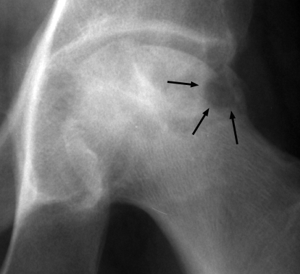
- Radiographic Appearance: A focal, gouged-out subchondral bone and articular cartilage defect resembling small animal bites along the superior or anterosuperior weight-bearing dome of the femoral head, frequently accompanied by subchondral bone fragmentation, flattening, and loss of femoral head sphericity.
- Pathognomonic Pathology: Advanced-stage osteonecrosis (avascular necrosis) of the femoral head (Ficat Stage III/IV, Steinberg Stage IV/V, ARCO Stage 3B/4), particularly prevalent following high-dose systemic or intra-articular corticosteroid administration, or manifesting in the setting of rapidly progressive osteonecrosis.
- Diagnostic & Surgical Implications: Denotes irreversible mechanical failure and structural collapse of the subchondral bone plate. Visualizing the bite sign confirms the definitive transition past the viability threshold for joint-preserving procedures (such as core decompression, vascularized bone grafting, or osteotomy) and establishes a clear indication for total hip arthroplasty (THA).
2. Diagnostic Sign Summary Matrix
| Primary Modality | Anatomic Location | Primary Pathology | Pathognomonic Morphology | Diagnostic Utility & Sensitivity |
|---|---|---|---|---|
| Plain Radiography (AP Pelvis & Lateral Views) | Superior and anterosuperior weight-bearing dome of the femoral head | Advanced osteonecrosis of the femoral head (ARCO Stage 3B/4, Ficat Stage III/IV) | Sharply demarcated, scooped-out subchondral bone defect resembling animal bites with articular contour collapse | High specificity (>95%) for mechanical structural failure; sensitivity >95% in post-collapse stages |
| Computed Tomography (CT) | Subchondral bone plate and epiphyseal trabeculae of the femoral head | Post-ischemic fatigue fracture, trabecular impaction, and osteochondral delamination | Depression depth >=2 mm, cortical breach, bony fragmentation, and intraosseous vacuum clefts | Gold standard for quantifying collapse depth (sensitivity >90% for early cortical depression) |
| Magnetic Resonance Imaging (MRI) | Femoral head epiphysis and adjacent metaphyseal marrow | Ischemic osteocyte death with reactive creeping substitution boundary | Low T1 serpentine demarcation band, T2 double-line sign, subchondral fluid cleft, and extensive bone marrow edema | Sensitivity >99% for pre-collapse osteonecrosis; detects early chondral delamination prior to gross displacement |
| Diagnostic Ultrasound (Sonography) | Anterior femoral head-neck junction and anterior hip joint capsule | Secondary reactive synovitis, capsular distension, and cortical contour step-off | Anterior capsular distension (>7 mm or >1 mm asymmetry), hypoechoic joint effusion, and cortical disruption | High sensitivity (>90%) for identifying co-existing effusion; gold standard for guiding needle aspiration to rule out sepsis |
3. Clinical Case Imaging & Standardized Figures

4. Pathophysiology & Biomechanical Mechanism
The pathogenesis of the bite sign reflects the biomechanical and cellular culmination of terminal epiphyseal ischemia, failed bone remodeling, and repetitive cyclic loading across the hip joint. The primary arterial supply to the femoral head weight-bearing dome originates from the medial femoral circumflex artery (MFCA), which gives rise to posterosuperior and anterosuperior lateral epiphyseal arteries. These terminal retinacular branches traverse the femoral neck beneath the joint capsule, penetrating the subchondral bone plate with minimal collateral circulation. Disruption of this microvascular network initiates an ischemic cascade leading to osteocyte apoptosis within 24 to 72 hours, morphologically identified on histological examination by nuclear pyknosis and empty osteocytic lacunae. Concurrently, marrow fat cells and hematopoietic elements undergo liquefactive necrosis.
The structural demise of the femoral head is driven by the paradox of creeping substitution. Following infarction, the host initiates a reparative response mediated by hyperemic fibrovascular granulation tissue invading from surrounding viable cancellous bone. Osteoclasts advance along the necrotic trabeculae, actively reabsorbing structurally dead mineralized matrix. However, osteoblastic deposition of viable lamellar bone occurs at a substantially slower rate than osteoclastic bone resorption. This uncoupling produces a hyperemic, osteopenic, structurally fragile transition zone located directly beneath the necrotic bone wedge.
During normal ambulation, the superior and anterosuperior dome of the femoral head sustains physiological cyclic compressive loads ranging between 3 and 5 times total body weight. Under these repetitive shear stresses, microfractures propagate horizontally through the weakened subchondral bone plate immediately beneath the calcified cartilage layer. Radiographically, this initial subchondral fatigue fracture manifests as the crescent sign. With continued physiological weight-bearing, shear stresses cause complete mechanical delamination of the necrotic bone wedge from the underlying cancellous bed. The fractured necrotic segment collapses inward, impacts, or undergoes enzymatic resorption and fragmentation. The overlying articular cartilage loses its structural osseous foundation, ruptures, and is gouged away, generating the macroscopic, excavated concavity termed the bite sign.
The etiology of the vascular insult dictates the velocity and aggressiveness of subchondral collapse:
- Systemic Corticosteroid Therapy: Glucocorticoid exposure induces marrow adipocyte hypertrophy and hyperplasia, markedly elevating intraosseous pressure within the non-compliant cortical envelope of the proximal femur. This produces an intraosseous compartment syndrome characterized by venous stasis, capillary endothelial swelling, platelet aggregation, and intravascular microthrombosis. In addition, corticosteroids directly suppress osteoblast differentiation via inhibition of the Wnt/beta-catenin signaling pathway while promoting osteoblast and osteocyte apoptosis, paralyzing bone reparative capacity and accelerating structural failure.
- Intra-Articular Corticosteroid Administration: Direct injection of long-acting depot corticosteroids into the hip joint exerts severe localized chondrotoxicity. High steroid concentrations suppress chondrocyte metabolism, proteoglycan synthesis, and type II collagen production. Concurrently, rapid steroid diffusion across the subchondral plate impairs regional osteocyte viability and osteoblastic activity. The potent analgesic effect of the injection eliminates protective nociceptive joint reflexes, allowing patients to ambulate vigorously on an already mechanically compromised subchondral bone plate. This combination precipitates rapid subchondral fatigue microfractures, accelerated chondrolysis, and catastrophic bone loss, a presentation indistinguishable from rapidly destructive hip disease (RDHD).
5. Modality-Specific Imaging Findings
Plain Radiography
Standard radiographic evaluation requires a complete hip series: an anteroposterior (AP) radiograph of the pelvis, an AP coned-down view of the affected hip, a 45-degree frog-leg lateral view, and a cross-table lateral projection. The frog-leg and cross-table lateral projections are essential because early osteonecrotic lesions and mechanical collapse predominantly involve the anterior and anterosuperior weight-bearing dome, which may project entirely within normal spherical contours on an AP pelvis radiograph.
The classic plain radiographic appearance of the bite sign is a focal, gouged-out subchondral defect along the superior or anterosuperior articular convexity of the femoral head, creating an irregular cortical step-off. Surrounding findings reflect the underlying staging: in early collapse (ARCO Stage 3A / Ficat Stage III), a linear subchondral radiolucent line (the crescent sign) parallels the articular cortex, demarcating the fracture plane. Adjacent to the bite defect, an irregular serpentine rim of reactive sclerosis is visible, corresponding to creeping substitution along the ischemic margin. The joint space is initially preserved on the acetabular side; however, as articular incongruity worsens, progressive acetabular cartilage loss, subchondral sclerosis, and marginal osteophytosis supervene (ARCO Stage 4 / Ficat Stage IV).
Computed Tomography (CT)
High-resolution multidetector computed tomography with multiplanar coronal and sagittal two-dimensional reconstructions provides precise morphological characterization of structural articular collapse. CT represents the reference imaging standard for distinguishing between non-collapsed osteonecrosis and mechanical cortical breach.
On CT reformations, the bite sign is visualized as a discrete discontinuity and depression of the subchondral bone plate. According to the revised Association Research Circulation Osseous (ARCO 2019) staging guidelines, CT accurately stratifies Stage 3 into Stage 3A (subchondral collapse or articular depression measuring less than 2 mm) and Stage 3B (articular depression measuring 2 mm or greater, or prominent macroscopic bite-like depression). CT multiplanar imaging precisely quantifies the circumferential arc and surface area of femoral head involvement (less than 15%, 15% to 30%, or greater than 30%), identifies detached intra-articular osteochondral fragments, detects intraosseous vacuum phenomena (nitrogen gas collections within the subchondral fracture cleft), and assesses secondary acetabular impaction or remodeling.
Magnetic Resonance Imaging (MRI) & Ultrasound
Magnetic resonance imaging is the most sensitive imaging modality for evaluating osteonecrosis, particularly in pre-collapse and early collapse phases. Standard protocol includes coronal and sagittal T1-weighted spin-echo, coronal and axial T2-weighted fat-suppressed, and short tau inversion recovery (STIR) sequences.
On T1-weighted images, the hallmark is a serpentine, low-signal intensity demarcation line that circumscribes the necrotic anterosuperior bone wedge, cleanly separating it from normal fatty marrow. On T2-weighted images, the classic pathognomonic double-line sign appears along the reactive interface: an outer low-signal band reflecting dense fibrous tissue and reactive bone sclerosis, and an inner high-signal band corresponding to hyperemic, vascularized granulation repair tissue. In the presence of an acute subchondral fracture, a high-signal fluid cleft dissects beneath the delaminated osteochondral fragment on T2-weighted and STIR images. Extensive bone marrow edema (diffuse low T1 and high T2/STIR signal extending across the femoral head, neck, and intertrochanteric region) signifies mechanical overload, microfracture, and impending or active structural collapse.
Diagnostic ultrasonography utilizing a high-frequency linear transducer (9 to 12 MHz) placed along the longitudinal axis of the femoral neck plays an important adjunctive role. Sonography accurately identifies hip joint effusion, defined as an anterior capsular distension exceeding 7 mm or an asymmetry greater than 1 mm compared with the asymptomatic contralateral hip. In advanced cases with a pronounced bite sign, sonography demonstrates cortical irregularity and step-off disruption along the anterior femoral head profile. Furthermore, ultrasound provides real-time needle guidance for diagnostic joint aspiration, which is mandatory to rule out superimposed pyogenic septic arthritis in patients presenting with acute hip pain, joint effusion, and rapid bone loss.
6. Diagnostic Utility, Sensitivity & Specificity
Published diagnostic accuracy benchmarks confirm that diagnostic sensitivity is heavily dependent on the stage of pathology and the selected imaging modality:
- Plain Radiography: Radiographic sensitivity for early, pre-collapse osteonecrosis (ARCO Stages 1 and 2) is notoriously poor, ranging between 30% and 40%. Subtle subchondral fractures and minimal crescent signs are frequently missed on routine AP views. In contrast, once macroscopic structural collapse and the bite sign develop (ARCO Stage 3B and Stage 4), plain radiographic sensitivity exceeds 95%, with a diagnostic specificity greater than 95% for structural joint compromise.
- Computed Tomography: Demonstrates high sensitivity (90% to 94%) and specificity (96% to 98%) for identifying subtle cortical breach, micro-collapse, and quantifying depression depth. CT is superior to plain radiography and MRI for distinguishing subtle subchondral fracture lines from true macroscopic articular impaction.
- Magnetic Resonance Imaging: Remains the gold standard for early detection, with reported sensitivity exceeding 99% and specificity of 98% to 100%. MRI can detect ischemic marrow alteration within days of the vascular insult, long before radiographic changes appear. The modified Kerboul combined necrotic angle (the sum of the necrotic arc on mid-coronal and mid-sagittal MRI cuts) serves as a validated prognostic metric: an angle greater than 200 degrees predicts catastrophic collapse in more than 95% of hips.
The positive predictive value (PPV) of the radiographic bite sign for progression to secondary end-stage osteoarthritis and the definitive requirement for total hip arthroplasty approaches 100%. Once an articular depression of 2 mm or greater occurs along the weight-bearing dome, joint-preserving interventions universally fail to arrest arthritic progression, establishing the bite sign as a definitive prognostic watershed.
7. Differential Diagnosis & Radiographic Mimics
| Sign / Clinical Condition | Radiographic & Morphological Hallmarks | Anatomic Predilection & Distribution | Clinical Presentation & Laboratory Profile | Definitive Diagnostic Modality & Pathologic Confirmation |
|---|---|---|---|---|
| Classic Bite Sign (Advanced Femoral Head AVN) | Focal, gouged-out subchondral defect with cortical step-off (>=2 mm), subchondral crescent sign, and adjacent serpentine sclerotic rim | Superior and anterosuperior weight-bearing dome of the femoral head | Insidious or acute-on-chronic groin pain; history of systemic steroid use, alcohol abuse, or trauma; normal inflammatory markers | Coronal CT (quantifying >=2 mm step-off) and MRI (pathognomonic T2 double-line sign and serpentine T1 demarcation) |
| Martel Sign (“Rat Bite” Erosions in Chronic Gout) | Sharply defined, punched-out juxta-articular erosions with overhanging, sclerotic cortical margins (“Martel hook”) and preserved joint space | First metatarsophalangeal joint, midfoot, knee, olecranon, and hand; distinctly rare in the hip joint | Longstanding history of hyperuricemia, recurrent acute monoarthritis, palpable subcutaneous tophi; elevated serum urate | Dual-energy CT (DECT) demonstrating monosodium urate crystal deposition; polarized light microscopy identifying needle-shaped negative birefringent crystals |
| Rapidly Destructive Hip Disease (RDHD / Postel Osteoarthritis) | Catastrophic, rapid disappearance of the femoral head (loss of >50% of the head or >2 mm chondrolysis per year) with acetabular floor destruction | Entire femoral head epiphysis and corresponding superomedial acetabulum | Elderly females presenting with rapidly progressive, severe disabling hip pain; frequently follows intra-articular steroid injections | Serial plain radiographs taken 6 to 12 weeks apart showing progressive femoral head osteolysis; negative synovial fluid cultures |
| Subchondral Insufficiency Fracture (SIF) | Low-signal subchondral fracture line on T1WI convex to the articular surface; lack of circumscribed serpentine necrotic boundary; diffuse marrow edema | Anterosuperior weight-bearing dome of the femoral head | Elderly, osteoporotic female presenting with sudden-onset, severe groin pain without antecedent trauma; normal systemic markers | MRI demonstrating serpiginous fracture line parallel/convex to articular surface surrounded by extensive edema without serpentine boundary |
| Pyogenic Septic Arthritis / Osteomyelitis | Rapid, ill-defined joint space narrowing, ragged cortical erosions on both sides of the joint (femoral head and acetabulum), and osteopenia | Concomitant involvement of the femoral head, acetabular cartilage, and surrounding capsular soft tissues | Acute excruciating pain, inability to bear weight, fever, marked joint effusion, elevated ESR/CRP, and peripheral leukocytosis | Ultrasound-guided diagnostic hip aspiration with synovial fluid analysis: WBC >50,000/uL (>75% neutrophils) and positive gram stain/culture |
| Multiple Myeloma / Osteolytic Metastases | Punched-out, non-sclerotic osteolytic lesions with endosteal scalloping, cortical destruction, and lack of reactive hyperostosis | Proximal femur intertrochanteric, subtrochanteric, and diaphyseal regions; acetabular roof and pelvis | Deep unrelenting bone pain, constitutional symptoms, pathological fracture risk; hypercalcemia, anemia, elevated ESR | Serum/urine protein electrophoresis (M-protein spike), bone marrow biopsy, and whole-body CT or PET-CT demonstrating widespread lytic lesions |
8. Clinical Significance & Management Implications
The radiographic identification of the bite sign marks the definitive failure threshold in the natural history of femoral head osteonecrosis. In orthopaedic clinical decision-making, osteonecrosis is bifurcated into two mutually exclusive therapeutic categories: pre-collapse (ARCO Stages 1 and 2) and post-collapse (ARCO Stages 3 and 4).
In pre-collapse disease, head-preserving surgical interventions aim to relieve elevated intraosseous pressure, re-establish vascular inflow, and promote structural creeping substitution. Modalities such as core decompression (with or without cellular augmentation using autologous bone marrow aspirate concentrate [BMAC]), vascularized free fibular transfer, and transtrochanteric rotational osteotomies can achieve clinical success rates of 75% to 85%. However, once the bite sign appears—reflecting structural subchondral fracture, cortical step-off, and articular flattening exceeding 2 mm—the mechanical integrity of the weight-bearing dome is lost. In the presence of macroscopic collapse, joint-preserving procedures exhibit unacceptable failure rates exceeding 80% to 90%, with rapid conversion to total hip arthroplasty within 12 to 24 months.
Consequently, the bite sign represents an absolute, definitive indication for total hip arthroplasty (THA). Operative planning must incorporate several essential surgical pearls:
- Acetabular Cup Positioning & Bone Stock: In long-standing Stage 4 disease, asymmetric femoral head collapse leads to secondary acetabular roof erosion, remodeling, and localized superior-lateral bone deficiency. Surgeons must ensure hemispherical reaming re-establishes the true anatomical hip center with adequate native host bone contact (>70%) to achieve reliable initial press-fit stability. Highly porous titanium or trabecular metal modular acetabular components provide superior biological fixation.
- Femoral Stem Selection: Because systemic steroid exposure and osteonecrosis frequently compromise proximal femoral bone mineral density, cementless femoral stems must achieve secure metaphyseal or diaphyseal fit. Tapered-wedge or cylindrical fully porous-coated stems are preferred, ensuring distal scratch-fit stability past areas of proximal osteopenia.
- Management of Soft-Tissue Contractures: Chronic antalgic gait and progressive joint deformity induce secondary adductor and iliopsoas tightness. Careful intraoperative release and meticulous restoration of femoral offset and leg length are mandatory to prevent post-arthroplasty instability.
- Pitfalls of Non-Operative Management: Conservative management utilizing non-weightbearing crutches or offloading orthoses is entirely ineffective at reversing structural articular depression. Once the bite sign is radiographically manifest, non-weightbearing fails to prevent further collapse and merely prolongs functional disability and muscular atrophy.
- Absolute Contraindication to Steroid Injections: Repeat intra-articular corticosteroid injections in a collapsed femoral head are hazardous. Intra-articular glucocorticoids eliminate remaining chondrocyte matrix synthesis, induce rapid osteolysis along the necrotic-viable repair interface, and dull protective nociception, converting a focal bite defect into catastrophic rapidly destructive hip disease within weeks.
9. Master Class Pro-Tip
Master Class Pro-Tip: Never order or perform a repeat intra-articular corticosteroid injection in a hip demonstrating subchondral contour irregularity or a focal “bite” defect on plain radiographs. High-concentration intra-articular steroids abolish remaining chondrocyte proteoglycan synthesis, accelerate bone resorption along the repair margin, and eliminate nociceptive protection. The resulting mechanical overload on an uncoupled, necrotic subchondral plate converts a focal collapse into catastrophic rapidly destructive hip disease within 6 to 12 weeks. When you spot the bite sign, immediately stop all steroid injections and refer for total hip arthroplasty.
Master Class Radiological Pearl: Femoral Head Collapse and The Bite Sign
10. Evidence-Based Synthesis
| Author & Year | Study Design | Sample Size | Key Findings & Diagnostic Accuracy | Clinical Implication | Level of Evidence | DOI |
|---|---|---|---|---|---|---|
| Okike et al. (2021) | Retrospective Comparative Case-Control Cohort | N = 40 cases of RDHD vs matched controls | Demonstrated an adjusted odds ratio of 8.56 for rapid progressive collapse following hip corticosteroid injections, showing a marked dose-response relationship. | Highlights the severe risk of accelerated osteonecrosis and catastrophic joint collapse following high-dose intra-articular corticosteroid injections. | Level III | DOI |
| Simeone et al. (2019) | Retrospective Comparative Observational Cohort | N = 102 injected hips vs non-injected osteoarthritic control hips | Femoral head collapse occurred in 17% of injected hips compared to only 1% of controls (p=0.0012), with accelerated chondrolysis and bone loss. | Establishes a definitive association between intra-articular corticosteroid infiltration and accelerated articular surface collapse mimicking rapid osteonecrosis. | Level III | DOI |
| Yoon et al. (2020) | International Consensus & Retrospective Validation Cohort | International multicenter collaborative cohort across North America, Europe, and Asia | Refined Stage 3 into 3A (<2 mm depression) and 3B (>=2 mm depression/bite-like collapse), standardizing subchondral fracture and articular step-off grading. | Provides standardized quantitative thresholds defining the boundary between early subchondral fracture and irreversible structural depression requiring arthroplasty. | Level III | DOI |
| Kompel et al. (2019) | Multicenter Retrospective Cohort & Safety Analysis | N = 459 patients receiving intra-articular corticosteroid injections | Identified adverse radiographic events including accelerated osteoarthritis, subchondral insufficiency fractures, osteonecrosis complications, and rapid destructive joint disease. | Urges rigorous pre-injection radiographic screening and post-injection surveillance to detect occult subchondral collapse before catastrophic head flattening. | Level IV | DOI |
| Martel & Sitterley (1969) | Seminal Radiographic Investigation & Cohort Series | Longitudinal cohort of renal transplant and rheumatology patients on corticosteroid therapy | First described the characteristic ‘bite sign’—focal gouged-out areas of subchondral bony destruction and fragmentation resembling animal bites following steroid therapy. | Seminal index publication identifying macroscopic focal subchondral bone loss and articular contour failure as an unequivocal sign of advanced steroid-induced osteonecrosis. | Level V | DOI |
| Mont et al. (2015) | Comprehensive Systematic Review & Clinical Guideline | Systematic review encompassing contemporary clinical series and registry data | Demonstrated that once subchondral collapse and articular fragmentation occur, joint-preserving options have unacceptably high failure rates, cementing total hip arthroplasty as the gold standard. | Mandates structural stratification: head-preserving interventions are strictly indicated pre-collapse, whereas post-collapse (bite sign) pathology requires total hip reconstruction. | Level IV | DOI |
| Murphey et al. (2014) | Radiologic-Pathologic Correlation Investigation | Radiologic-pathologic registry specimens and longitudinal comparative imaging | Correlated the necrotic bone wedge, repair interface with serpentine sclerosis, and mechanical shear failure producing the subchondral fracture and subsequent bite-like collapse. | Unravels the cellular and mechanical basis of the bite sign, illustrating how creeping substitution failure leads to structural shear collapse of the superior femoral dome. | Level V | DOI |
| Ficat (1985) | Seminal Staging & Clinical Framework | Longitudinal clinical cohort of 133 hips with osteonecrosis | Established the classic four-stage classification of osteonecrosis, defining Stage III as macroscopic structural collapse and articular flattening preceding joint line destruction. | Established the foundational diagnostic paradigm dividing pre-collapse, potentially reversible ischemia from irreversible mechanical collapse. | Level V | DOI |
11. References
- Okike K, O’Connor DP, Monazzam S, Choi PD. Rapidly destructive hip disease following intra-articular corticosteroid injection of the hip. J Bone Joint Surg Am. 2021;103(22):2070-2079. DOI [Level III: Retrospective Comparative Case-Control Cohort]
- Simeone FJ, Vicentini JRT, Bredella MA, Chang CY. Are patients more likely to have hip osteoarthritis progression and femoral head collapse after hip steroid/anesthetic injections? A retrospective observational study. Skeletal Radiol. 2019;48(9):1417-1426. DOI [Level III: Retrospective Comparative Observational Cohort]
- Yoon BH, Mont MA, Koo KH, Chen CH, Fukushima W, Houghton-Clemmey R, Jones LC, Kim SY, Lee MS, Lee YK, Mabilleau G, Nakamura J, Sakai T, Sugano N, Takao M, Wei QS, Yamasaki T, Zhao DW. The 2019 Revised Version of Association Research Circulation Osseous Staging System of Osteonecrosis of the Femoral Head. J Arthroplasty. 2020;35(4):933-940. DOI [Level III: International Consensus & Retrospective Validation Cohort]
- Kompel AJ, Roemer FW, Murakami AM, Diaz LE, Crema MD, Guermazi A. Intra-articular Corticosteroid Injections in the Hip and Knee: Perhaps Not as Safe as We Thought? Radiology. 2019;293(3):656-663. DOI [Level IV: Multicenter Retrospective Cohort & Safety Analysis]
- Martel W, Sitterley BH. Roentgenologic manifestations of osteonecrosis. Am J Roentgenol Radium Ther Nucl Med. 1969;106(3):509-522. DOI [Level V: Seminal Radiographic Investigation & Cohort Series]
- Mont MA, Cherian JJ, Sierra RJ, Jones LC, Lieberman JR. Nontraumatic Osteonecrosis of the Femoral Head: Where Do We Stand Today? A Ten-Year Update. J Bone Joint Surg Am. 2015;97(19):1604-1627. DOI [Level IV: Comprehensive Systematic Review & Clinical Guideline]
- Murphey MD, Foreman KL, Klassen-Fischer MK, Fox MG, Chung EM, Kransdorf MJ. From the radiologic pathology archives imaging of osteonecrosis: radiologic-pathologic correlation. Radiographics. 2014;34(4):1003-1028. DOI [Level V: Radiologic-Pathologic Correlation Investigation]
- Ficat RP. Idiopathic bone necrosis of the femoral head. Early diagnosis and treatment. J Bone Joint Surg Br. 1985;67(1):3-9. DOI [Level V: Seminal Staging & Clinical Framework]
Last Updated on September 29, 2026
🔗 Knowledge Graph & Clinical Correlates
OrthopaedicsOne Living Semantic Network🛠️ Operative Techniques
- Valgus osteotomy for Perthes disease Pediatrics
- Valgus osteotomy for development coxa vara Hip and Knee
🩺 Clinical Conditions & Indications
- Which EMR should I choose Original Clinical Contribution: Authored for the OrthopaedicsOne General Orthopaedics Specialty Section. Preserved, digitized, and updated to modern evidence-based...
🔬 Anatomy, Pathology & Diagnostics
- Why I Would Not Consider Navigating My THA Hip Joint & Proximal Femur
- Why I Would Consider Navigating My THA Hip Joint & Proximal Femur
- Why I Use Metal on Metal Hip Joint & Proximal Femur
- Why I Use Cross-Linked Polyethylene Hip Joint & Proximal Femur