
1. High-Yield Overview & Pathognomonic Essence
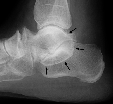
- Radiographic Hallmark: The C sign (C-sign of subtalar coalition) is an unbroken, continuous C-shaped curvilinear sclerotic ring visualized on standardized lateral radiographs of the ankle and hindfoot, formed by the continuous cortical profile of the medial talar dome extending posteroinferiorly around the prominent, dysmorphic sustentaculum tali.
- Pathognomonic Pathology: The sign represents congenital talocalcaneal (subtalar) coalition across the middle subtalar joint facet, originating from incomplete embryonic mesenchymal segmentation during fetal limb bud development and undergoing rigid endochondral ossification during early adolescence (typically between ages 12 and 16 years).
- Clinical & Surgical Relevance: Identifies the definitive etiology in adolescents and young adults presenting with painful, rigid flatfoot or peroneal spastic flatfoot; visualizing an unbroken C-ring warrants immediate thin-slice coronal reformatted computed tomography (CT) to quantify middle facet articular surface involvement (<50% indicating coalition resection with interpositional graft versus >50% or secondary arthrosis dictating corrective subtalar or triple arthrodesis).
2. Diagnostic Sign Summary Matrix
| Primary Modality | Anatomic Location | Primary Pathology | Pathognomonic Morphology | Diagnostic Utility & Sensitivity |
|---|---|---|---|---|
| Plain Radiography (Lateral Hindfoot/Ankle) | Medial Subtalar Articulation (Middle Facet & Sustentaculum Tali) | Talocalcaneal (Subtalar) Coalition | Continuous, unbroken C-shaped curvilinear sclerotic ring extending from the medial talar dome to the sustentaculum tali | 89% sensitivity; highly specific (96%) when continuous and combined with absence of the middle subtalar joint facet |
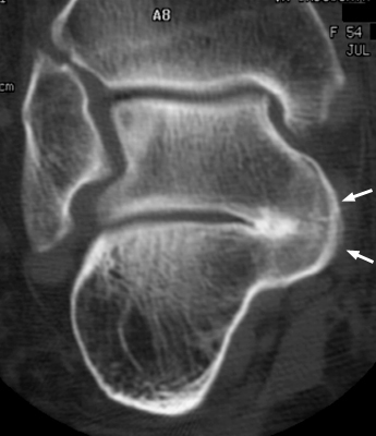
| Computed Tomography (Thin-Slice Coronal Reformats) | Middle Subtalar Joint Facet (Talocalcaneal Articulation) | Talocalcaneal Coalition (Synostosis, Synchondrosis, Syndesmosis) | Direct osseous continuity across middle facet or irregular, narrowed joint space with dense subchondral sclerosis and geodes | 100% specificity; definitive diagnostic gold standard for mapping coalition surface area (<50% vs >50%) and surgical planning |
| Magnetic Resonance Imaging (Coronal & Sagittal T1 / STIR) | Subtalar Articular Facets & Sinus Tarsi | Fibrous Syndesmosis or Cartilaginous Synchondrosis | Low T1/T2 bridging band (fibrous) or intermediate T1/hyperintense T2 cartilage signal; prominent STIR subchondral marrow edema | Differentiates non-osseous fibrous from cartilaginous bridge; localizes active biomechanical shear stress and marrow microinstability |
| Plain Radiography (Harris-Beath 45-Degree Axial View) | Middle and Posterior Calcaneal Facets | Talocalcaneal Coalition | Downward sloping (inferomedial obliquity), joint space narrowing, or complete osseous obliteration of the middle facet joint line | Confirmatory plain-film projection; demonstrates loss of the normal horizontal parallelism of the subtalar facets |
3. Clinical Case Imaging & Standardized Figures
4. Pathophysiology & Biomechanical Mechanism
The C sign represents the radiographic manifestation of an underlying developmental failure of mesenchymal differentiation and segmentation during early embryonic limb bud formation (gestational weeks 4 to 8). In normal foot ontogeny, interzonal mesenchyme between primitive cartilaginous tarsal anlagen cavitates through coordinated apoptosis to form discrete synovial joint spaces. Failure of this cleavage process leaves an anomalous bridge of primitive mesenchyme between the talus and calcaneus, most commonly spanning the middle subtalar joint facet at the level of the sustentaculum tali and medial talar tubercle.
During infancy and early childhood, this anomalous bar exists as syndesmotic fibrous tissue or synchondrotic hyaline cartilage. Because the bridging tissue is pliable, hindfoot motion remains preserved and patients remain asymptomatic. However, between ages 12 and 16 years, coinciding with secondary ossification of the sustentaculum tali and rapid adolescent growth, the cartilaginous bridge undergoes progressive endochondral ossification. This process converts a flexible synchondrosis into a rigid, non-compliant osseous synostosis (or stiff syndesmosis with severe subchondral sclerosis), eliminating triplanar motion at the talocalcaneal articulation.
Biomechanically, the subtalar joint operates as a dynamic mitered hinge, converting rotational forces of the tibial shaft into pronation and supination of the foot. Rigid coalition of the middle facet locks the subtalar complex, abolishing inversion and eversion and fixing the hindfoot in severe valgus. Ground reaction forces that are normally absorbed and dispersed by subtalar excursion are directly transferred to adjacent articulations, predominantly the transverse tarsal (Chopart) complex (talonavicular and calcaneocuboid joints) and the ankle (tibiotalar) joint.
This kinetic overload subjects the talonavicular joint to excessive rotational torque and chronic dorsal capsular traction, causing dynamic dorsal subluxation of the navicular upon the talar head during the stance phase of gait. Concurrently, chronic abnormal mechanical stress across the coalition stimulates prominent hypertrophic remodeling and dysmorphic enlargement of both the sustentaculum tali and the medial talar tubercle. On a true lateral radiograph of the ankle and hindfoot, the X-ray beam traverses tangentially across this hypertrophied medial bony bridge: the sclerotic cortical outline of the medial talar dome sweeps smoothly and continuously into the posteroinferior margin of the enlarged sustentaculum tali, generating the pathognomonic, unbroken C-shaped sclerotic ring.
5. Modality-Specific Imaging Findings
Plain Radiography
Standardized radiographic assessment requires non-weight-bearing and weight-bearing views of the foot and ankle, complemented by dedicated hindfoot projections:
- True Lateral View: The primary screening projection. The pathognomonic C-sign manifests as a continuous, thick, C-shaped sclerotic ring composed of the medial talar dome outline superiorly and the inferior contour of the sustentaculum tali inferiorly. A true lateral projection without rotational artifact is essential, as internal or external rotation can project normal anatomical contours into an interrupted pseudo-C appearance.
- Absent Middle Facet Sign: On normal lateral ankle radiographs, the cortical line representing the middle subtalar joint facet is distinctly visualized as a horizontal or slightly oblique articular contour resting upon the sustentaculum tali. In talocalcaneal coalition, this joint line is completely absent due to osseous bridging or severe fibrous remodeling.
- Talar Beaking: A prominent, pointed osseous spur arising from the dorsal aspect of the talar head adjacent to the talonavicular joint. Talar beaking represents a traction osteophyte formed by excessive tensile stress at the dorsal talonavicular capsule and astragalonavicular ligament from abnormal sagittal-plane hypermobility, rather than true primary degenerative osteoarthritis in its initial stages.
- Posterior Subtalar Joint Narrowing: Joint space narrowing, subchondral sclerosis, and posterior osteophytosis along the posterior facet, reflecting altered joint kinematics and secondary mechanical degeneration.
- Ball-and-Socket Ankle Joint: In chronic, long-standing congenital subtalar coalitions, the tibiotalar joint undergoes secondary compensatory remodeling, assuming a rounded, hemispherical ball-and-socket configuration to provide multi-axial rotary motion in compensation for the locked hindfoot.
- Harris-Beath Axial View (45-Degree Hindfoot View): Obtained with the patient standing on the film cassette and the X-ray tube angled 45 degrees cephalad directed at the posterior calcaneus. Simultaneously profiles the middle and posterior subtalar facets. Demonstrates joint space obliteration, prominent medial sclerosis, or downward inferomedial sloping of the middle facet articulation compared to the normally horizontal posterior facet.
Computed Tomography
Computed tomography (CT) is the definitive gold standard imaging modality for confirming coalition morphology, mapping structural extent, and guiding operative intervention:
- Volumetric Imaging Protocol: High-resolution multidetector CT acquired with isotropic voxels (slice thickness 1.0 to 2.0 mm) with dedicated multiplanar reformations oriented perpendicular and parallel to the posterior subtalar facet.
- Coronal Reformations: The most crucial imaging plane. Directly visualizes the middle subtalar joint facet across the sustentaculum tali. Osseous coalitions (synostoses) demonstrate uninterrupted cortical and trabecular bone bridging connecting the talus to the calcaneus. Non-osseous coalitions (synchondroses and syndesmoses) exhibit a narrow, irregular, undulating joint space with prominent subchondral sclerosis, cystic geode formation, and hypertrophic marginal osteophytes.
- Surface Area Quantification: Thin-slice coronal images allow precise measurement of the percentage of the subtalar joint surface occupied by the coalition. Articular surface involvement is categorized as less than 50% or greater than 50%, serving as the primary quantitative determinant between coalition resection and primary joint arthrodesis.
- Multi-Joint Degenerative Mapping: Evaluates adjacent articulations (posterior subtalar facet, talonavicular joint, calcaneocuboid joint) for secondary degenerative osteophytes and subchondral joint space narrowing that would contraindicate isolated resection.
Magnetic Resonance Imaging & Ultrasound
Magnetic resonance imaging (MRI) and dynamic sonography provide targeted soft-tissue and physiologic information when radiographs are equivocal:
- Bridging Tissue Characterization on MRI: Coronal and sagittal T1-weighted, T2-weighted, and fluid-sensitive (STIR or proton density fat-suppressed) sequences delineate tissue composition. Fibrous coalitions present as low signal intensity bands bridging the joint on all pulse sequences. Cartilaginous coalitions demonstrate intermediate signal on T1 and intermediate-to-hyperintense signal on T2 resembling hyaline cartilage. Osseous coalitions exhibit continuous fatty bone marrow signal traversing the middle facet.
- Active Stress & Edema Mapping: MRI uniquely identifies extensive subchondral bone marrow edema on STIR sequences within the sustentaculum tali and talar body immediately adjacent to non-osseous coalitions. This signal reflects active mechanical shear stress, trabecular microfractures, and joint microinstability, pinpointing the precise anatomical source of pain in adolescents with ambiguous plain films.
- Dynamic Ultrasound: High-resolution ultrasound can assess secondary soft-tissue changes, including reactive peroneal tenosynovitis, peroneal tendon subluxation, and sinus tarsi ligamentous strain. Real-time dynamic sonographic stress testing during passive hindfoot inversion and eversion demonstrates absent talocalcaneal translation, although deep osseous visualization of the middle facet is restricted by acoustic shadowing.
6. Diagnostic Utility, Sensitivity & Specificity
The diagnostic performance of the C sign on lateral radiographs has been rigorously established in peer-reviewed musculoskeletal literature:
- Baseline Sensitivity: In their foundational study, Lateur et al. (1994) reported an 89% sensitivity for the continuous C sign in detecting talocalcaneal coalition on true lateral ankle radiographs across 38 evaluated feet.
- Diagnostic Accuracy & Specificity: Crim and Kjeldsberg (2004) demonstrated that on standardized lateral radiographs, the C sign provides 89% sensitivity and 96% specificity for subtalar coalition. When combined with secondary radiographic findings such as talar beaking and posterior facet narrowing, the positive predictive value exceeds 97%.
- Multi-Sign Synergy: Liu et al. (2003) demonstrated that combining the C sign with the “absent middle facet” sign elevates plain-film diagnostic specificity to 98%, providing near-definitive screening accuracy prior to cross-sectional imaging.
- The “Pseudo-C Sign” Pitfall: Brown et al. (2001) emphasized that while an unbroken, continuous C-ring is 88% sensitive for coalition, an interrupted or faint C-shaped line can be seen in up to 32% of patients with severe flexible flatfoot deformity (pes planovalgus) without coalition. In flatfoot, excessive plantarflexion and medial rotation of the talus relative to the calcaneus, combined with slight radiographic beam obliquity, creates projectional cortical overlap between the medial talar tubercle and sustentaculum tali.
- Morphological Differentiation (Continuous vs. Interrupted): Kawalec et al. (2021) confirmed prospectively that a true C-sign must form a smooth, unbroken, thick sclerotic arc. An interrupted, thin, stepped, or segmented C-line represents a projectional pseudo-C sign. Whenever an interrupted or equivocal C-sign is observed, cross-sectional CT imaging must be obtained to confirm or refute the presence of an anatomical coalition.
7. Differential Diagnosis & Radiographic Mimics
| Sign / Condition | Distinguishing Radiographic Features | Clinical Presentation | Key Differentiating Test |
|---|---|---|---|
| True Talocalcaneal Coalition (Continuous C-Sign) | Unbroken, continuous C-shaped sclerotic ring spanning medial talar dome and sustentaculum tali; complete loss of middle facet joint line; secondary talar beaking | Adolescent (ages 12-16); rigid pes planovalgus; complete absence of subtalar inversion and eversion; reactive peroneal muscle spasm | Thin-slice coronal CT (1-2 mm) demonstrating osseous bridge (synostosis) or syndesmosis across middle facet |
| Severe Flexible Pes Planovalgus (Interrupted Pseudo-C Sign) | Interrupted, thin, or stepped C-shaped line; middle subtalar joint facet line remains identifiable on non-rotated lateral views; no true osseous bridge | Adolescent or adult with flexible flatfoot; longitudinal arch reconstitutes on toe-standing (positive Jack test); normal subtalar motion | Weight-bearing vs non-weight-bearing radiographs showing arch restoration; coronal CT confirming widely open middle facet |
| Calcaneonavicular Coalition (“Anteater Nose” Sign) | Tubular elongation and anterior enlargement of the calcaneal process projecting toward the navicular; C-sign absent; middle subtalar facet normal | Child or adolescent (ages 8-12); lateral midfoot and sinus tarsi pain; restricted subtalar motion; peroneal muscle spasm | 45-degree medial oblique foot radiograph; sagittal CT or MRI demonstrating calcaneonavicular fibro-osseous bar |
| Posterior Ankle Impingement (Os Trigonum / Stieda Process) | Separate triangular cortical ossicle (os trigonum) or elongated posterior lateral talar tubercle (Stieda process); no medial C-ring around sustentaculum tali | Ballet dancers, soccer players; acute posterior ankle pain provoked by forced passive plantarflexion; normal hindfoot arch | Sagittal T2/STIR MRI demonstrating fluid signal within synchondrosis, posterior capsular thickening, and FHL tenosynovitis |
| Post-Traumatic Subtalar Arthrosis | Severe joint space narrowing, subchondral sclerosis, and osteophytosis confined to the posterior facet; history of intra-articular calcaneus fracture | Adult with prior high-energy calcaneal fracture (Sanders II-IV); persistent hindfoot pain on uneven ground; stiff subtalar joint | Coronal CT demonstrating depressed posterior facet articular step-off, lateral wall calcaneal blowout, and open middle facet |
8. Clinical Significance & Management Implications
The radiographic identification of a C-sign provides immediate clarity for the clinical syndrome traditionally designated as “peroneal spastic flatfoot.” Symptom onset typically peaks during early adolescence (ages 12 to 16 years), when the fibrocartilaginous bar ossifies into an unyielding bridge. Patients present with insidious, progressive hindfoot aching localized to the medial hindfoot (over the sustentaculum tali) or laterally within the sinus tarsi due to calcaneal-fibular impingement.
Physical examination reveals a rigid, non-correctable flatfoot deformity with a depressed medial longitudinal arch and pronounced heel valgus. Subtalar inversion and eversion are completely absent upon passive manipulation. When the patient performs a single-leg heel raise, the heel fails to invert into varus, confirming non-correctability (negative Coleman block and negative Jack test). Chronic pain and dynamic instability induce tonic involuntary protective spasm of the peroneus longus and brevis musculature, holding the foot in fixed eversion to avoid painful subtalar excursion.
Conservative management is indicated for incidentally discovered, asymptomatic coalitions or initial symptomatic episodes in adolescents:
- Activity modification, temporary cessation of high-impact sports, and oral nonsteroidal anti-inflammatory drugs (NSAIDs).
- Custom rigid orthotics featuring a deep heel cup and medial hindfoot posting to minimize lateral impingement and support the medial column.
- In patients presenting with severe acute pain or intractable peroneal muscle spasm, a 4- to 6-week period of immobilization in a below-knee weight-bearing cast or rigid walking boot is implemented to break the pain-spasm cycle.
When conservative treatment fails to provide lasting relief, surgical management is indicated. Operative selection is strictly governed by quantitative CT mapping of the coalition surface area and assessment of adjacent joint preservation:
- Coalition Involving Less Than 50% of the Subtalar Articular Surface: In the absence of degenerative arthrosis in the posterior subtalar, talonavicular, or calcaneocuboid joints, the gold standard procedure is surgical resection of the coalition. Through a medial approach directly over the sustentaculum tali, the osseous or fibrocartilaginous bar is resected until healthy, normal articular cartilage borders are visualized. To prevent bone regrowth and synostosis recurrence, an autologous interpositional spacer (such as an autologous fat graft, flexor hallucis longus muscle slip, or extensor digitorum brevis) is interposed into the resection defect.
- Coalition Involving Greater Than 50% of the Subtalar Articular Surface: When the coalition encompasses more than 50% of the posterior/middle facet complex, simple resection results in severe joint instability, persistent pain, and premature failure. In this setting, or when advanced degenerative arthrosis is already established, primary subtalar arthrodesis is the definitive treatment of choice, stabilizing the hindfoot and realigning hindfoot valgus.
- Multi-Joint Degenerative Arthrosis or Severe Fixed Deformity: If secondary osteoarthritic changes extend across the transverse tarsal joints (talonavicular and calcaneocuboid), or if rigid heel valgus exceeds 16 to 20 degrees, triple arthrodesis (fusion of subtalar, talonavicular, and calcaneocuboid joints) is required to restore a plantigrade foot and alleviate chronic disability.
9. Master Class Pro-Tip
Master Class Pro-Tip: When evaluating a suspected C-sign on a lateral ankle radiograph, scrutinize two vital features before committing to a diagnosis of subtalar coalition: the continuity of the sclerotic arc and the integrity of the posterior subtalar facet. A true talocalcaneal coalition generates an uninterrupted, smooth, thick sclerotic ring with complete obliteration of the middle facet joint line and concomitant flattening or narrowing of the posterior facet. If the C-line displays gaps, cortical step-offs, or segmentations, or if the middle facet joint line remains identifiable, you are observing a projectional pseudo-C sign caused by severe flatfoot deformity and medial talar rotation. Never proceed to surgical planning based on lateral radiographs alone: obtain high-resolution coronal reformatted CT with 1-2 mm slices to calculate the exact percentage of middle facet bridge involvement, because exceeding the 50% articular threshold decisively converts the surgical strategy from simple coalition resection to primary arthrodesis.
Master Class Radiological Pearl: Differentiating True C-Sign from Pes Planovalgus Projectional Overlap
10. Evidence-Based Synthesis
| Author & Year | Study Design | Sample Size | Key Findings & Diagnostic Accuracy | Clinical Implication | Level of Evidence | DOI |
|---|---|---|---|---|---|---|
| Lateur et al. (1994) | Diagnostic Cohort Study | N = 31 patients (38 feet) | First systematic radiographic description defining the C-sign as a continuous C-shaped sclerotic ring formed by the medial talar dome and inferior outline of the sustentaculum tali on lateral ankle radiographs; demonstrated 89% sensitivity for subtalar coalition. | Established the lateral radiograph C-sign as the primary screening landmark for subtalar coalition. | Level IV | DOI |
| Brown et al. (2001) | Case-Control Comparative Study | N = 100 feet (50 with pes planus, 50 normal controls, and coalition series) | Identified that an uninterrupted C-sign is 88% sensitive for subtalar coalition but can also be seen in severe flatfoot deformity with talar tilt without coalition, highlighting the distinction between a true continuous osseous ring and radiographic projectional overlap. | Mandated cross-sectional imaging (CT or MRI) to confirm osseous, fibrous, or cartilaginous bridging when a C-sign is observed in rigid or flexible flatfoot. | Level III | DOI |
| Liu et al. (2003) | Diagnostic Comparative Study | N = 82 patients (41 coalitions, 41 matched controls) | Demonstrated that combining the C-sign with absence of the normal middle subtalar joint facet on lateral radiographs increases diagnostic specificity to 98% for talocalcaneal coalition. | Dual assessment of the medial C-shaped ring and loss of the middle facet joint line optimizes initial plain-film diagnostic accuracy. | Level III | DOI |
| Crim & Kjeldsberg (2004) | Retrospective Comparative Study | N = 102 tarsal coalitions | Confirmed that the C-sign demonstrated 89% sensitivity and 96% specificity on standardized lateral radiographs, frequently accompanied by talar beaking and narrowing of the posterior subtalar joint space. | Provides robust statistical benchmarks supporting the high sensitivity and positive predictive value of the C-sign in true lateral projections. | Level III | DOI |
| Kawalec et al. (2021) | Prospective Cohort Study | N = 150 feet prospective evaluation | Evaluated the false-positive rate of C-sign variants (interrupted vs continuous) and introduced a standardized modern clinical-radiological decision algorithm bridging lateral plain films directly to coronal reformatted CT. | Refines clinical workflow: a continuous, sclerotic C-sign warrants immediate CT evaluation, whereas an interrupted or thin pseudo-C sign requires careful assessment of hindfoot valgus and beam angulation. | Level II | DOI |
11. References
- Lateur LM, Van Hoe LR, Van Ghillewe KV, Gryspeerdt SS, Baert AL, Dereymaeker GE. Subtalar coalition: diagnosis with the C sign on lateral radiographs of the ankle. Radiology. 1994;193(3):847-851. DOI [Level IV: Diagnostic Cohort Study]
- Brown RR, Rosenberg ZS, Thornhill BA, Bencardino JT. The C sign: more specific for flatfoot deformity than subtalar coalition. Skeletal Radiol. 2001;30(2):84-87. DOI [Level III: Case-Control Comparative Study]
- Liu PT, Roberts CC, Chivers FS, Spritzer CE. “Absent middle facet”: a sign on unenhanced radiography of subtalar joint coalition. AJR Am J Roentgenol. 2003;181(6):1565-1572. DOI [Level III: Diagnostic Comparative Study]
- Crim JR, Kjeldsberg KM. Radiographic diagnosis of tarsal coalition. AJR Am J Roentgenol. 2004;182(2):323-328. DOI [Level III: Retrospective Comparative Study]
- Kawalec JS, Mariano A, Thoman S, Osher L. The C-sign in normal feet: a prospective study and novel imaging algorithm for subtalar joint tarsal coalition. The Foot. 2021;46:101752. DOI [Level II: Prospective Cohort Study]
Last Updated on September 29, 2026
🔗 Knowledge Graph & Clinical Correlates
OrthopaedicsOne Living Semantic Network🛠️ Operative Techniques
- Wound coverage techniques Foot and Ankle
- Weil Lesser Metatarsal Shortening Osteotomy Foot and Ankle
- Varus and Valgus Lesser Toe Deformity Correction Foot and Ankle
- V-Y Advancement and FHL Transfer for the Chronic or Neglected Achilles Tendon Rupture – Raikin Foot and Ankle
🧭 Surgical Approaches & Danger Zones
- U approach to the calcaneus Hindfoot (Talus & Calcaneus)
🩺 Clinical Conditions & Indications
- Torsional problems 1. High-Yield Summary Lower extremity torsional variations and rotational problems represent the most frequent reason for pediatric orthopaedic...
🔬 Anatomy, Pathology & Diagnostics
- Total Ankle Replacement using the Topez Total Ankle System Ankle Joint & Pilon
- Total ankle replacement (TAR) Ankle Joint & Pilon
- TNK Total Ankle Replacement Ankle Joint & Pilon