
1. High-Yield Overview & Pathognomonic Essence
The crowded carpal sign is a classic, highly specific radiographic indicator of high-energy perilunate disruption, alerting clinicians to gross midcarpal dislocation on standard posteroanterior (PA) wrist radiographs:
- Fundamental Radiographic Appearance: Manifests on the posteroanterior (PA) view of the wrist as abnormal overlapping and crowding of the distal carpal row (capitate and hamate) over the proximal carpal row (lunate and triquetrum), with obliteration of normal intercarpal joint spaces, severe foreshortening of carpal height, and disruption of Gilula arcs I and II.
- Pathognomonic Pathology: Represents the frontline radiographic hallmark of volar perilunate dislocation (and severe complex perilunar instability patterns), reflecting complete ligamentous disengagement at the midcarpal joint through the space of Poirier with proximal migration of the distal carpal row.
- Critical Diagnostic & Surgical Implication: Serves as an immediate clinical red flag for an orthopaedic emergency, commanding urgent closed reduction, mandatory evaluation for acute median nerve compression within the carpal tunnel, and definitive open reduction and internal fixation (ORIF) with ligamentous reconstruction.
2. Diagnostic Sign Summary Matrix
| Primary Modality | Anatomic Location | Primary Pathology | Pathognomonic Morphology | Diagnostic Utility & Sensitivity |
|---|---|---|---|---|
| Plain Radiography (PA View) | Midcarpal and Radiocarpal Joint Columns | Volar Perilunate Dislocation | Abnormal overlapping of the distal carpal row onto the proximal row; foreshortened carpus; disruption of Gilula arcs I and II | High specificity (>95%) for perilunate disruption; primary screening sign on initial emergency plain radiograph series |
| Plain Radiography (Lateral View) | Radius-Lunate-Capitate Longitudinal Column | Volar Perilunate Dislocation | Lunate maintains normal seating within the distal radial lunate fossa while the capitate head displaces volarly (palmarward) | 100% specific on true lateral projection; definitively differentiates volar perilunate dislocation from classic lunate dislocation |
| Computed Tomography (CT) | Carpal Skeleton & Articular Surfaces | Perilunate Fracture-Dislocation (Lesser vs. Greater Arc) | Multiplanar mapping of volar capitate dislocation, osteochondral shearing fractures, and greater arc fracture lines | Gold standard for operative planning; >95% sensitivity for occult fractures (scaphoid, capitate, triquetrum) and loose bodies |
| Magnetic Resonance Imaging (MRI) | Intrinsic & Extrinsic Carpal Ligaments; Median Nerve | Ligamentous Disruption & Median Nerve Impingement | Discontinuity of scapholunate (SLIL) and lunotriquetral (LTIL) ligaments; tear of space of Poirier; median nerve compression | Definitive soft-tissue assessment; detects early avascular necrosis (AVN) of proximal pole of scaphoid and lunate; evaluates carpal tunnel neuropathy |
3. Clinical Case Imaging & Standardized Figures
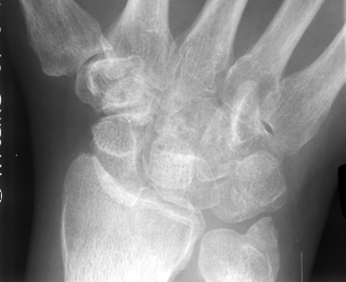
Detailed analysis of Figure 1 reveals the classic morphological hallmarks of the crowded carpal sign on a standard posteroanterior (PA) radiograph of the wrist. The physiological 2 mm intercarpal joint space separating the proximal and distal carpal rows is completely obliterated. The proximal pole of the capitate and the adjacent hamate have migrated proximally into the space normally occupied by the lunate and triquetrum, creating dense osseous superimposition. In addition, Gilula carpal arcs I and II exhibit marked step-off deformities and discontinuity, and the overall carpal height is markedly foreshortened, confirming gross carpal architectural collapse characteristic of a high-energy volar perilunate dislocation.
4. Pathophysiology & Biomechanical Mechanism
The crowded carpal sign is the two-dimensional radiographic manifestation of a three-dimensional catastrophic mechanical failure of the carpus. Under physiologic conditions, carpal stability is maintained through an intricate balance of osseous geometry, intrinsic interosseous ligaments, and robust extrinsic capsular ligaments. The proximal carpal row (scaphoid, lunate, and triquetrum) functions as an intercalated segment lacking direct tendinous insertions, moving passively between the forearm and the rigid distal carpal row (trapezium, trapezoid, capitate, and hamate).
Whereas dorsal perilunate dislocations (accounting for greater than 97% of perilunate injuries) typically result from extreme wrist hyperextension, ulnar deviation, and intercarpal supination as delineated by Mayfield and colleagues, volar perilunate dislocations (representing less than 3% of cases) arise through distinctly different force vectors. Volar perilunate injuries are produced by violent wrist hyperflexion combined with axial loading, high-energy axial compression coupled with sudden intercarpal pronation, or secondary anterior translation during an unstable reduction attempt of a dorsal perilunate pattern.
Central to the pathomechanics of perilunate dislocation is the failure of the space of Poirier. The space of Poirier is an anatomically vulnerable, ligament-free area within the volar carpal capsule located between the distally converging radioscaphocapitate (RSC) ligament laterally and the long radiolunate (LRL) ligament medially, situated directly superficial to the lunocapitate articulation. When severe torsional and shear stresses overload the wrist, this capsule tears widely. In volar perilunate dislocations, midcarpal disengagement occurs as the proximal articular pole of the capitate escapes anteriorly across the volar lip of the lunate cup.
Once the lunocapitate articulation is disrupted and the scapholunate interosseous ligament (SLIL) and lunotriquetral interosseous ligament (LTIL) are ruptured, the rigid architectural tethering of the midcarpal joint is eliminated. The distal carpal row is subjected to the unopposed, powerful longitudinal traction of the extrinsic forearm musculature—including the flexor digitorum profundus, flexor digitorum superficialis, flexor carpi radialis, and the wrist extensors. This longitudinal pull drives the distal carpal row proximally into the carpal tunnel and into the anatomical space normally reserved for the proximal row.
On a posteroanterior radiograph, this proximal migration causes the distal carpal bones (predominantly the capitate and hamate) to superimpose directly over the lunate, triquetrum, and scaphoid. This produces marked carpal foreshortening, reflected by a precipitous drop in the Youm carpal height ratio (normally 0.54 ± 0.03) to values well below 0.45. Concurrently, the smooth, congruent contours of Gilula arcs I and II are disrupted: Arc III (the proximal contour of the capitate and hamate) overlaps and obliterates Arc II (the distal contour of the proximal row), producing the dense, congested, “crowded” silhouette pathognomonic of the sign.
5. Modality-Specific Imaging Findings
Plain Radiography
Plain radiography represents the frontline diagnostic modality in all acute wrist trauma. A standard trauma series must include neutral posteroanterior (PA), true lateral, and semi-pronated oblique views:
- Posteroanterior (PA) View: Demonstrates the definitive crowded carpal sign. The normal 2 mm joint spaces of the midcarpal and radiocarpal joints are obliterated. The distal carpal row overrides the proximal row, creating an increased radiographic density from double-cortical superimposition. Gilula arc I (proximal articular margin of scaphoid, lunate, and triquetrum) and arc II (distal concave margin of the proximal row) demonstrate obvious step-offs, breaks, or complete non-visualization. The scaphoid frequently displays a “cortical ring sign” (signet ring sign) due to foreshortening in acute palmar flexion, or exhibits a visible fracture line through its waist in trans-scaphoid variants.
- Lateral View: The lateral radiograph is the decisive projection to establish carpal column collinearity and differentiate perilunate from lunate dislocation. On a true lateral radiograph (where the distal radial articular surface is perpendicular and the pisiform projects over the central third of the scaphoid waist), the lunate maintains its normal concentric articulation within the lunate fossa of the distal radius (preserved radiolunate articulation). In sharp contrast, the capitate head and distal carpal row are displaced anteriorly (volarly) to the lunate. This relationship is the exact opposite of dorsal perilunate dislocation (where the capitate is displaced dorsally) and distinct from lunate dislocation (where the lunate is enucleated volarly into the carpal tunnel while the capitate remains aligned with the radius).
- Oblique Projections: Helpful in unmasking subtle avulsion fractures of the radial styloid, dorsal triquetral ridge, or capitate neck, which often accompany high-energy ligamentous failure.
Computed Tomography (CT)
Multi-detector CT with thin-slice (0.5 mm to 1.0 mm) acquisitions and multiplanar (coronal, sagittal, and axial) as well as 3D volume-rendered reconstructions is essential following initial closed reduction:
- Fracture Mapping (Lesser vs. Greater Arc): CT precisely categorizes the injury pattern into lesser arc (purely ligamentous perilunate disruption) or greater arc injuries (trans-scaphoid, trans-radial styloid, trans-capitate, or trans-triquetral fracture-dislocations). Sagittal reconstructions map the exact fracture geometry, displacement, and comminution of scaphoid waist or capitate fractures.
- Articular Congruency & Osteochondral Shearing: Coronal and sagittal slices clearly visualize intra-articular shearing fragments, particularly avulsions from the dorsal or volar lips of the lunate and capitate head, which frequently become entrapped in the midcarpal joint and mechanically impede anatomical closed reduction.
- Carpal Tunnel & DRUJ Architecture: Axial CT reconstructions quantify the degree of carpal tunnel volumetric narrowing and evaluate for associated subluxation of the distal radioulnar joint (DRUJ) or fracture of the hook of the hamate.
Magnetic Resonance Imaging (MRI) & Ultrasound
While CT remains the primary advanced osseous modality in acute trauma, MRI and point-of-care ultrasound offer specialized diagnostic capabilities:
- Magnetic Resonance Imaging (MRI): High-resolution 3T MRI utilizing T1-weighted, T2-weighted fat-suppressed, and proton density-weighted sequences provides direct visualization of the intrinsic interosseous ligaments. Complete discontinuity of the dorsal band of the scapholunate interosseous ligament (SLIL) and lunotriquetral interosseous ligament (LTIL) is readily confirmed. Extrinsic capsular disruption, particularly through the space of Poirier and the radioscaphocapitate ligament, is mapped. Furthermore, T1-weighted hypointensity and extensive bone marrow edema patterns in the scaphoid proximal pole and lunate provide early prognostic data regarding compromised intraosseous vascular perfusion and risk of avascular necrosis (AVN).
- Diagnostic Ultrasound: In the emergency resuscitation bay, high-frequency linear ultrasound can rapidly assess acute median nerve compression within the carpal tunnel. The anteriorly displaced carpal row creates marked flattening of the median nerve against the transverse carpal ligament, with a cross-sectional area frequently swelling beyond 12 mm² at the carpal tunnel inlet, accompanied by tense subretinacular hematoma.
6. Diagnostic Utility, Sensitivity & Specificity
Perilunate dislocations represent notorious diagnostic traps in emergency and orthopaedic practice. In a landmark multicenter investigation of 166 perilunate injuries, Herzberg and colleagues documented that up to 25% of perilunate dislocations and fracture-dislocations are missed or delayed on initial emergency room evaluation. Diagnostic delays directly correlate with poor functional outcomes, persistent median nerve neuropathy, progressive carpal collapse, and accelerated post-traumatic osteoarthritis.
The crowded carpal sign provides an indispensable, frontline screening safeguard. When present on a standard posteroanterior radiograph, carpal overlapping and disruption of Gilula arcs exhibit a diagnostic specificity exceeding 95% to 98% for major carpal dislocation or high-grade instability. However, the diagnostic sensitivity of the PA view alone ranges between 70% and 75% in general triage settings. Inexperienced observers frequently attribute subtle carpal overlap to technical positioning error or obliquity, rather than recognizing true proximal migration.
When the PA radiograph is evaluated systematically alongside a true lateral view, diagnostic sensitivity approaches 98% to 100%. The positive predictive value (PPV) of the crowded carpal sign in the setting of acute, painful wrist trauma with severe functional restriction exceeds 95%. Technical pitfalls leading to diagnostic failure include reliance on rotated or oblique “pseudo-lateral” radiographs, failure to trace Gilula arcs systematically, omission of carpal height ratio measurement, and presence of distracting polytrauma such as severe head injury or ipsilateral long-bone fractures.
7. Differential Diagnosis & Radiographic Mimics
Systematic comparison of the crowded carpal sign with other major carpal dislocations and traumatic instability patterns is essential for prompt and accurate management:
| Sign / Condition | Frontal (PA) Radiographic Features | Lateral Radiographic Key Relationships | Distinguishing Hallmarks & Gold Standard |
|---|---|---|---|
| Volar Perilunate Dislocation (Crowded Carpal Sign) | Prominent carpal crowding and overlap; Gilula arcs I & II disrupted; reduced carpal height ratio (<0.45) | Lunate maintains normal seating within distal radius lunate fossa; capitate head displaced volarly (palmarward) | True lateral view establishes collinear radius-lunate axis with anterior capitate displacement; very high risk of acute carpal tunnel syndrome |
| Dorsal Perilunate Dislocation (Classic Mayfield III) | Disrupted Gilula arcs I & II; foreshortened carpal height; scaphoid ring sign or scaphoid waist fracture | Lunate maintains normal articulation with distal radius; capitate head displaced dorsally relative to lunate cup | Most common perilunate dislocation pattern (>97%); lateral radiograph demonstrates dorsal displacement of the capitate column |
| Lunate Dislocation (Mayfield Stage IV) | Triangular “piece-of-pie sign” due to rotated, palmar-flexed lunate; disrupted Gilula arcs I & II | Capitate aligns with distal radius axis; lunate enucleated and tilted volarly into carpal tunnel (“spilled teacup sign”) | End-stage perilunar instability; capitate drops into radial fossa while lunate is pushed anteriorly into the carpal tunnel |
| Midcarpal Dislocation / Dynamic Instability | Normal or near-normal carpal height; subtle midcarpal step-offs; absent gross carpal bone overlap | Abnormal scapholunate angle (>70° in DISI, <30° in VISI); lunate tilted dorsally or volarly without gross midcarpal dislocation | Dynamic stress radiography and fluoroscopy unmask midcarpal click; absence of gross continuous overlapping on static PA views |
| Axial Carpal Dislocation (Axial-Radial / Axial-Ulnar) | Widening between longitudinal carpal columns; carpus split into two divergent halves; metacarpal splaying | Disruption of both radiocarpal and midcarpal columns; severe soft-tissue blast or crush injury | High-energy crush trauma; marked widening between carpal columns on PA view rather than isolated proximal-distal crowding |
| Trans-scaphoid Perilunate Fracture-Dislocation | Crowded carpal sign present; displaced fracture through scaphoid waist with proximal scaphoid pole staying with lunate | Proximal scaphoid fragment and lunate align with radius; distal scaphoid fragment and capitate displace volarly or dorsally | Greater arc injury (De Quervain injury pattern); multiplanar CT confirms scaphoid waist fracture line and fragment alignment |
8. Clinical Significance & Management Implications
Identification of the crowded carpal sign dictates immediate, aggressive orthopaedic intervention. Perilunate dislocations are true surgical emergencies that carry devastating morbidity if managed conservatively or if diagnosis is delayed:
- Emergency Closed Reduction: Urgent closed reduction must be performed in the emergency setting without delay under adequate intravenous sedation, regional block, or general anesthesia. Longitudinal traction is applied using fingertraps with 10 to 15 pounds of countertraction for 10 to 15 minutes to fatigue the spastic forearm musculature. For a volar perilunate dislocation, reduction involves maintaining longitudinal traction with the wrist in slight extension, applying direct dorsal-to-palmar manual pressure over the displaced distal carpus, and gently flexing the wrist to guide the capitate head back over the volar lip of the lunate into the lunate cup. Immediate post-reduction neurovascular assessment and confirmatory orthogonal radiographs are mandatory.
- Urgent Carpal Tunnel Decompression: Volar perilunate dislocations carry an exceptionally high incidence of acute median nerve neuropraxia and acute compartment syndrome of the carpal tunnel. The anteriorly translated distal carpal row and associated hematoma directly crowd the carpal canal, compressing the median nerve against the rigid transverse carpal ligament. Any persistent digital paresthesia, two-point discrimination loss, or progressive motor deficit mandates emergent formal open carpal tunnel release (CTR).
- Definitive Operative Reconstruction: Nonoperative management or prolonged closed immobilization leads to unacceptably high rates of recurrent subluxation, secondary carpal collapse, and disabling arthrosis (failure rate exceeding 60% to 70%). Definitive open reduction and internal fixation (ORIF) with ligamentous repair is the gold standard of care.
- Surgical Approaches & Stabilization: A combined dorsal and volar approach is widely regarded as the most robust strategy. The volar approach facilitates extended carpal tunnel release, direct median nerve inspection, evacuation of hematoma, and anatomic suture anchor repair of the torn space of Poirier and volar radiocarpal ligaments (radioscaphocapitate and long radiolunate). The dorsal approach (via the 3rd or 4th extensor compartment with dorsal capsulotomy) provides direct visualization of the radiocarpal and midcarpal joints, enables extraction of osteochondral fragments, ensures anatomic reduction of the lunocapitate and scapholunate joints, and allows direct suture anchor repair of the dorsal scapholunate (SLIL) and lunotriquetral (LTIL) ligaments. Trans-fixation with multiple 1.4 mm or 1.6 mm Kirschner wires across the scapholunate (2 wires), lunotriquetral, and lunocapitate joints provides rigid temporary neutralization. Associated scaphoid waist fractures undergo rigid headless compression screw fixation.
- Postoperative Rehabilitation: Patients are immobilized in a thumb spica cast for 8 to 10 weeks, followed by Kirschner wire removal and a structured hand therapy regimen emphasizing protected range of motion and progressive strengthening.
9. Master Class Pro-Tip
Master Class Pro-Tip: Whenever a frontal PA wrist radiograph reveals carpal crowding with overlapping silhouettes and loss of Gilula arcs, never accept an oblique or rotated lateral view. Demand a true, cross-table lateral radiograph with the radial styloid superimposed over the pisiform. Immediately scrutinize the collinear alignment of the radius, lunate, and capitate: if the lunate remains squarely seated in the radial fossa while the capitate head is displaced palmarward, you have confirmed a volar perilunate dislocation; if the lunate is tipped palmarward like a spilled teacup while the capitate remains aligned with the radius, it is a lunate dislocation. Remember that volar perilunate dislocations exert massive direct anterior pressure on the median nerve—test sensation in the digital pulp immediately before and after reduction, and maintain an ultra-low threshold for emergency open carpal tunnel release.
Master Class Radiological Pearl: Volar Perilunate Dislocation and Carpal Crowding
10. Evidence-Based Synthesis
| Author & Year | Study Design | Sample Size | Key Findings & Diagnostic Accuracy | Clinical Implication | Level of Evidence | DOI |
|---|---|---|---|---|---|---|
| Klein & Webb (1987) | Clinical Case Series & Radiographic Description | Cohort series | First described the crowded carpal sign on frontal wrist radiographs, characterized by abnormal overlapping of the distal carpal row over the proximal carpal row due to proximal migration of the capitate and distal carpal row during volar perilunate dislocation. | Pathognomonic indicator of volar perilunate dislocation on frontal PA radiographs, alerting clinicians to gross carpal disruption even when lateral views are suboptimal or misinterpreted. | Level IV | DOI |
| Gilula (1979) | Radiological Investigation & Classification | N = 100 wrist injury cases | Established the three smooth carpal arcs (Gilula arcs) on PA wrist radiographs; demonstrated that disruption, step-off, or overlap of any arc reliably indicates ligamentous disruption, fracture, or carpal subluxation/dislocation. | Standardized frontline radiological evaluation of wrist trauma; disruption of Arcs I and II with overlapping carpal silhouettes confirms perilunate injury. | Level IV | DOI |
| Mayfield et al. (1980) | Biomechanical Cadaveric Investigation | N = 32 fresh-frozen cadaveric wrists | Delineated 4 progressive stages of perilunar instability (scapholunate dissociation -> lunocapitate disruption -> lunotriquetral disruption -> lunate extrusion into carpal tunnel) under hyperextension, ulnar deviation, and intercarpal supination/pronation vectors. | Provided the foundational anatomic and biomechanical classification of perilunate and lunate dislocations, explaining the sequential failure of carpal ligaments that permits distal row displacement. | Level V | DOI |
| Herzberg et al. (1993) | Multicenter Retrospective Cohort Study | N = 166 perilunate injuries | Found that up to 25% of perilunate dislocations and fracture-dislocations are missed or delayed on initial emergency radiographic evaluation, directly correlating with adverse long-term functional scores and secondary post-traumatic arthritis. | Emphasizes the vital clinical importance of recognizing subtle PA signs such as the crowded carpal sign and carpal arc step-offs to prevent catastrophic diagnostic delays. | Level III | DOI |
| Stanbury & Elfar (2011) | Comprehensive Clinical & Surgical Review | Systematic expert synthesis | Synthesized diagnostic algorithms, acute closed reduction maneuvers, emergency carpal tunnel decompression criteria, and combined dorsal-volar open reduction internal fixation (ORIF) and ligamentous reconstruction protocols. | Confirms that perilunate dislocations are true orthopaedic emergencies requiring urgent anatomic reduction and stable ligamentous restoration to prevent avascular necrosis and median nerve neuropathy. | Level V | DOI |
| Panagopoulos et al. (2017) | Case Report & Systematic Review of Volar Dislocations | N = 1 (Review of 22 published volar perilunate cases) | Demonstrated that volar perilunate dislocations account for <3% of all perilunate dislocations; confirmed that the crowded carpal sign on PA radiograph together with volar capitate displacement on lateral radiograph provides definitive diagnostic confirmation. | Reinforces awareness of rare volar perilunate variants which present with profound carpal crowding and high rates of acute median nerve compression requiring urgent open decompression. | Level V | DOI |
| Bhat et al. (2019) | Clinical Case Investigation & Literature Review | N = 1 (Review of volar perilunate patterns) | Characterized the complex injury biomechanics involving hyperflexion and axial loading leading to palmar displacement of the carpus with extensive carpal overlap on frontal projections. | Highlights the mandatory role of preoperative multiplanar CT in mapping occult osteochondral shearing fractures and guide surgical approach selection. | Level V | DOI |
11. References
- Klein A, Webb LX. The crowded carpal sign in volar perilunar dislocation. J Trauma. 1987;27(1):82-84. DOI [Level IV: Clinical Case Series]
- Gilula LA. Carpal injuries: analytic approach and case exercises. AJR Am J Roentgenol. 1979;133(3):503-517. DOI [Level IV: Radiological Investigation]
- Mayfield JK, Johnson RP, Kilcoyne RK. Carpal dislocations: pathomechanics and progressive perilunar instability. J Hand Surg Am. 1980;5(3):226-241. DOI [Level V: Biomechanical Investigation]
- Herzberg G, Comtet JJ, Linscheid RL, Amadio PC, Cooney WP, Stalder J. Perilunate dislocations and fracture-dislocations: a multicenter study. J Hand Surg Am. 1993;18(5):768-779. DOI [Level III: Multicenter Retrospective Cohort Study]
- Stanbury SJ, Elfar JC. Perilunate dislocation and perilunate fracture-dislocation. J Am Acad Orthop Surg. 2011;19(9):554-562. DOI [Level V: Comprehensive Review]
- Panagopoulos A, Bougiouklis T, Prelorentzos G, Grigoropoulos P, Kokkalis Z, Chronopoulos E. Volar trans-scaphoid perilunate dislocation of the wrist: a case report. Med Sci Case Rep. 2017;4:65-70. DOI [Level V: Case Report & Systematic Review]
- Bhat AK, Kurup H, Acharya A. Volar translunate perilunate dislocation: case report and analysis of review of literature. J Hand Microsurg. 2019;11(Suppl 1):S15-S19. DOI [Level V: Case Report & Literature Review]
Last Updated on September 28, 2026
🔗 Knowledge Graph & Clinical Correlates
OrthopaedicsOne Living Semantic Network🛠️ Operative Techniques
- Wrist Implant Arthroplasty Hand and Wrist
- Wrist disarticulation Hand and Wrist
- Wrist Dennervation Hand and Wrist
- Wrist block Pain Management
🧭 Surgical Approaches & Danger Zones
- Volar approach to the wrist Wrist and Carpus
- Volar approach to the scaphoid Wrist and Carpus
🩺 Clinical Conditions & Indications
- Wrist sprains 1. High-Yield Summary Wrist sprain is an imprecise clinical descriptor that frequently obscures high-energy ligamentous disruption of the...
- Wrist fractures Introduction Summary about significance of fracture including epidemiology. Anatomy Review the relevant anatomy of the bone, the joint,...
- Wartenberg’s syndrome 1. High-Yield Summary Wartenberg's syndrome (cheiralgia paresthetica) is an entrapment neuropathy of the superficial branch of the radial...
- Volar plate injury 1. High-Yield Summary Volar plate injuries of the proximal interphalangeal (PIP) joint represent the most frequent soft-tissue and...
🔬 Anatomy, Pathology & Diagnostics
- Wrist joint Wrist and Carpus
- Watson test Wrist and Carpus