
1. High-Yield Overview & Pathognomonic Essence
- Morphological Hallmark: The Cupid’s bow contour is characterized on frontal (anteroposterior) radiographs of the lumbar spine by paired, upward-curving arcuate concavities along the inferior vertebral endplate separated by a central triangular bony ridge, which project on lateral radiographs as a single smooth, localized depression along the posterior two-thirds of the inferior endplate.
- Normal Developmental Variant: It represents a benign, non-pathological anatomical variant of vertebral endochondral ossification and annular Sharpey fiber anchorage occurring predominantly at the L4 and L5 levels (and occasionally L3), found in approximately 10% to 14% of the population, with normal bone mineral density and intact trabecular architecture.
- Critical Diagnostic Utility: Immediate recognition of this “do-not-touch” contour prevents catastrophic misdiagnosis of acute or osteoporotic vertebral compression fractures, traumatic depressed endplate fractures, Schmorl’s nodes, or osteolytic neoplastic collapse, thereby averting unwarranted spinal immobilization, bone biopsy, antiresorptive pharmacological escalation, or unnecessary cement augmentation (vertebroplasty/kyphoplasty).
2. Diagnostic Sign Summary Matrix
| Primary Modality | Anatomic Location | Primary Pathology | Pathognomonic Morphology | Diagnostic Utility & Sensitivity |
|---|---|---|---|---|
| Plain Radiography (Frontal AP & Lateral Views) | Inferior endplate of lower lumbar vertebrae (predominantly L4 and L5; less commonly L3) | Normal developmental anatomical variant (asymptomatic endochondral remodeling) | AP view: Paired symmetrical parasagittal concavities with central downward-pointing bony promontory; Lateral view: Superimposed smooth posterior inferior endplate depression | High specificity (>98%) when characteristic bidirectional morphology is recognized; plain film sensitivity approximately 10% to 14%, directly reflecting population prevalence |
| Computed Tomography (Multiplanar Axial & 2D Coronal/Sagittal Reformations) | Posterior two-thirds of inferior lumbar vertebral endplates (L3-L5) | Benign developmental cortical remodeling at annular attachment zones | Smooth, bilobed symmetrical endplate depressions with intact, non-disrupted, well-corticated subchondral margins; normal cancellous trabeculae without fracture lines, sclerosis, or retropulsion | Gold standard for definitive confirmation of cortical integrity (>99% specificity); definitively rules out cortical fragmentation, intraosseous vacuum clefts, or osseous destruction |
| Magnetic Resonance Imaging (Sagittal & Coronal T1, T2, & STIR Sequences) | Lower lumbar vertebral bodies, endplates, and adjacent intervertebral disc spaces | Normal physiological bone marrow architecture and preserved disc mechanics | Normal homogeneous marrow signal on all pulse sequences with complete absence of bone marrow edema (no STIR hyperintensity); preserved disc hydration and height without intraosseous herniation | 100% negative predictive value for acute traumatic/osteoporotic fracture or infection; distinguishes quiescent developmental contour from acute inflammatory or malignant endplate collapse |
3. Clinical Case Imaging & Standardized Figures
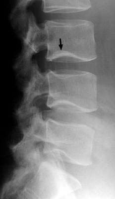

4. Pathophysiology & Biomechanical Mechanism
The Cupid’s bow contour represents an entirely benign, physiological developmental variant of vertebral endochondral ossification, rather than an acquired pathological deformity or osteopenic microfracture. During adolescent spinal growth and skeletal maturation, the primary ossification center of the vertebral body centrum expands radially toward the peripheral ring apophyses and cartilaginous endplates. In the lower lumbar spine, where the lordotic curvature and axial gravitational loading generate high biomechanical shear stresses, focal variations in the rate and geometry of endochondral bone formation naturally occur.
Specifically, the contour arises from a localized, bilateral parasagittal developmental depression along the posterior two-thirds of the inferior vertebral endplate. At these specific parasagittal zones, the outer annular lamellae of the intervertebral disc (fibers of Sharpey) insert with exceptional mechanical anchorage into the cartilaginous endplate and subchondral bone. During the period of rapid spinal lengthening in late childhood and adolescence, relative focal deceleration of endochondral ossification beneath these robust annular insertions creates two symmetrical, smooth, upward-arcing concavities. Between these paired depressions, a central midline ridge or promontory of mature cortical and cancellous bone persists, forming the cephalad-pointing peak of the classical Cupid’s bow.
Historically, early radiological theories hypothesized that the Cupid’s bow contour might represent focal osteopenia, persistent notochordal remnants, or subclinical microtrabecular insufficiency collapse. However, landmark imaging-histopathologic and densitometric investigations by Chan, Sartoris, Resnick, and colleagues (1997) decisively refuted these assumptions. In their correlation of vertebral specimens and prospective patient cohorts, dual-energy X-ray absorptiometry (DEXA) demonstrated that vertebral bodies exhibiting the Cupid’s bow contour maintain identical volumetric and areal bone mineral density compared to unaffected vertebrae, with no evidence of focal osteopenia.
Histopathological analysis confirmed that the subchondral bone beneath the concavities consists of a continuous, intact, and well-organized cartilaginous plate supported by thickened cortical lamellar bone. The underlying cancellous trabeculae demonstrate robust architecture without microfractures, reactive callus formation, osteonecrosis, or fibrovascular granulation tissue. Dense arrays of Sharpey fibers seamlessly anchor into the intact bone, proving that the contour reflects a permanent, harmonious adaptation between native ligamentous-annular insertions and skeletal endochondral remodeling.
From a biomechanical standpoint, the Cupid’s bow contour preserves normal vertebral stiffness, compressive load distribution, and segmental compliance. Because the subchondral plate remains continuous and structurally reinforced by thickened cortical bone, the vertebral body resists axial compression without focal vulnerability. The regional morphology accommodates physiological disc deformation during flexion, extension, and axial rotation without predisposing the motion segment to accelerated degeneration, disc herniation, or structural instability.
5. Modality-Specific Imaging Findings
Plain Radiography
Plain radiography represents the primary imaging modality where the Cupid’s bow contour is initially encountered, typically as an incidental finding. Accurate diagnostic interpretation requires mandatory correlation between the frontal (anteroposterior) and lateral projections:
- Anteroposterior (AP) Projection: Demonstrates the pathognomonic appearance. The inferior endplate of L4, L5, or L3 presents paired, smooth, upward-curving arcuate radiolucent concavities situated symmetrically on either side of the midline. A central triangular bony promontory or keel points cephalad between the two scallops, completing the classic silhouette of a drawn Cupid’s bow. The subchondral cortex along the entire contour is thin, continuous, and sharply demarcated, without cortical interruption, ragged borders, or surrounding reactive osteosclerosis.
- Lateral Projection: Because the paired concavities are anatomically confined to the posterior two-thirds of the inferior endplate, the X-ray beam traverses both depressions in profile. On the lateral radiograph, these bilateral scallops superimpose precisely, projecting as a single, smooth, localized concavity or depression in the posterior half of the inferior endplate. Crucially, the anterior third to half of the inferior endplate remains straight and flat, and the anterior vertebral body height is completely preserved without wedging, step-off, or cortical buckling.
Computed Tomography
Computed tomography (CT) provides definitive cross-sectional confirmation of the benign developmental nature of the sign, resolving any diagnostic ambiguity created by overlapping bowel gas or pelvic structures on plain radiographs:
- Axial CT: Depicts two well-circumscribed, bilobed, kidney-shaped or crescentic depressions located symmetrically within the posterior half of the inferior endplate. The cortex lining the depressions is intact, smooth, and uniformly thin, without jagged fracture lines, cortical step-offs, or defect margins. The adjacent subchondral trabecular bone shows normal attenuation (Hounsfield units) without focal osteopenia, trabecular impaction bands, intraosseous gas (vacuum clefts), or retropulsed bone fragments within the spinal canal.
- Coronal Reformations: Faithfully reproduce the frontal plain film appearance, clearly demonstrating the paired upward-arcing subchondral scallops and the central bony ridge, confirming that the cortex is completely intact across the entire transverse width of the endplate.
- Sagittal Reformations: Demonstrate the localized concavity confined to the posterior inferior endplate, verifying that the superior endplate, anterior cortex, and posterior vertebral wall maintain normal geometric alignment and cortical integrity.
Magnetic Resonance Imaging & Ultrasound
Magnetic resonance imaging (MRI) is the definitive modality for confirming normal marrow physiology and excluding acute or destructive pathology:
- T1-Weighted Sequences: Demonstrates completely normal, homogeneous signal intensity matching adjacent non-affected lumbar vertebrae, representing physiological adult fatty and hematopoietic marrow. A thin, uninterrupted hypointense line marks the intact subchondral bone cortex. There is no band-like hypointensity or marrow replacement.
- T2-Weighted and STIR (Short Tau Inversion Recovery) Sequences: Displays absolute absence of bone marrow edema. The subchondral spongiosa shows normal signal without any hyperintense inflammatory edema or fluid. The absence of STIR hyperintensity carries a 100% negative predictive value for acute traumatic fractures, acute osteoporotic insufficiency fractures, active spondylodiscitis, or neoplastic marrow infiltration.
- Intervertebral Disc & Soft Tissues: Disc height and hydration within the adjacent intervertebral space are fully preserved. There is no intraosseous herniation of nucleus pulposus through a disrupted endplate (distinguishing it from an acute Schmorl’s node). The anterior and posterior longitudinal ligaments are intact, and there is no prevertebral soft tissue edema or epidural mass.
- Ultrasound: Ultrasound has no role in the adult lumbar spine due to acoustic shadowing from the overlying bony pelvis and posterior elements. In neonatal and infant high-frequency spinal sonography, unossified cartilaginous vertebral endplates can be evaluated, showing smooth contours without hematoma, cortical step-offs, or cartilaginous clefts.
6. Diagnostic Utility, Sensitivity & Specificity
The primary clinical value of the Cupid’s bow contour lies in its status as an established, highly specific normal anatomical variant. Epidemiological and radiological cohort studies demonstrate that the contour is present in approximately 10% to 14% of the population (Dietz & Christensen 1976, Ramirez et al. 1984). Anatomically, it occurs with marked predilection for the lower lumbar spine, presenting most frequently at L4 (approximately 60% to 70% of identified cases), followed by L5 (25% to 35%), and only occasionally at L3 (5% to 10%). It is exceptionally rare in the upper lumbar or thoracic spine.
When the characteristic bidirectional imaging features are appreciated, plain radiography and computed tomography demonstrate a diagnostic specificity exceeding 98% to 99% for benign developmental anatomy. Because it is a normal congenital variant, sensitivity parameters reflect population prevalence rather than disease detection. In prospective longitudinal pediatric cohorts, such as the Canadian STOPP Consortium National Pediatric Bone Health Study (Jaremko et al. 2015), the Cupid’s bow contour was identified as one of the most frequent normal developmental variants emerging during adolescent skeletal growth that is mistakenly classified as a vertebral compression fracture in children undergoing bone fragility evaluations.
The paramount diagnostic pitfall associated with the Cupid’s bow contour is false-positive overdiagnosis. On isolated lateral plain radiographs, the superimposed posterior concavity can be misinterpreted by clinicians unfamiliar with the variant as an acute depressed endplate fracture, a fish-mouth biconcave osteoporotic deformity, or an erosive lesion. Conversely, on isolated AP views, the radiolucent parasagittal scallops can be misconstrued as osteolytic metastases or infectious subchondral destruction. Recognizing the mutual correspondence of the AP and lateral projections immediately resolves these diagnostic dilemmas.
7. Differential Diagnosis & Radiographic Mimics
| Sign / Condition | Distinguishing Radiographic Features | Clinical Presentation | Key Differentiating Test |
|---|---|---|---|
| Cupid’s Bow Contour (Normal Variant) | Paired parasagittal smooth concavities on AP view with central bony ridge; superimposed single posterior concavity on lateral view; intact, non-disrupted subchondral cortex; preserved anterior height | Asymptomatic incidental finding or incidental to non-specific mechanical low back pain; normal neurovascular examination | Multiplanar CT confirms smooth, intact cortication; MRI demonstrates normal marrow without STIR edema (100% specificity) |
| Osteoporotic Vertebral Compression Fracture (OVF) | Loss of anterior or middle vertebral body height; sharp cortical step-off, anterior cortex buckling, or impaction band of condensed trabeculae; biconcave deformity crossing the entire endplate width | Acute or subacute focal axial back pain, often following minimal trauma or ordinary daily activities; elderly, postmenopausal females, or chronic corticosteroid therapy | MRI displays band-like T1 hypointensity and intense STIR bone marrow hyperintensity during acute/subacute phases; CT reveals trabecular impaction and cortical disruption |
| Traumatic Depressed Endplate Fracture | Jagged cortical breach, sharp comminuted fracture lines extending into the vertebral body, focal endplate depression; potential retropulsion of bone fragments into the spinal canal | Acute severe focal back pain immediately following moderate-to-high energy trauma (fall from height, motor vehicle collision); possible localized tenderness and neurological deficit | Multiplanar CT clearly delineates jagged cortical disruption, comminution, and canal retropulsion; MRI demonstrates diffuse acute marrow edema |
| Schmorl’s Node / Scheuermann’s Disease | Focal, well-circumscribed, round or irregular central endplate defect with surrounding sclerotic rim; Scheuermann’s exhibits multiple contiguous thoracic/thoracolumbar levels with anterior wedging (>5 degrees across 3+ vertebrae) and kyphosis | Typically asymptomatic incidental finding; acute localized pain during initial acute disc extrusion; adolescent thoracic hyperkyphosis in Scheuermann’s disease | Sagittal MRI demonstrates focal herniation of disc material into the vertebral spongiosa with surrounding edema in the acute phase; CT shows a circumscribed focal crater rather than broad paired scallops |
| Pyogenic Spondylodiscitis | Ragged, irregular, ill-defined destructive erosions involving contiguous endplates across the intervertebral disc; severe loss of disc space height; prevertebral and epidural soft tissue phlegmon or abscess | Severe, constant, unremitting spinal pain, exquisite tenderness to percussion, constitutional symptoms (fever, chills, night sweats), elevated ESR and CRP | Contrast-enhanced MRI demonstrates disc and bivertebral T2/STIR hyperintensity, hyperintense fluid within the collapsed disc, and rim-enhancing epidural or paraspinal abscesses |
| Lytic Metastasis / Multiple Myeloma | Ill-defined, “moth-eaten” or permeative osteolytic destruction; cortical thinning and pathological cortical breach; involvement of posterior elements (pedicle destruction, “winking owl” sign); pathologic collapse without sclerotic borders | Progressive, dull, unremitting nocturnal or rest pain; history of primary malignancy (breast, lung, prostate, renal, thyroid) or systemic myeloma symptoms; systemic weight loss and fatigue | Contrast-enhanced MRI shows discrete focal masses with low T1, high T2/STIR signal, and marked contrast enhancement; PET-CT or CT-guided percutaneous core needle biopsy provides definitive cytology |
8. Clinical Significance & Management Implications
The Cupid’s bow contour carries profound clinical and medicolegal importance as a classic “do-not-touch” non-fracture deformity. Because it is a permanent, entirely benign developmental variation, its identification requires absolute clinical restraint, patient reassurance, and zero medical, radiological, or surgical intervention. Differentiating this variant from acute or pathologic spinal conditions is critical to protecting patients from iatrogenic harm, unnecessary invasive procedures, and financial burdens.
Foremost, correct recognition prevents the misdiagnosis of acute osteoporotic vertebral compression fractures. In elderly or osteopenic patients presenting with non-specific lower back pain, an incidental Cupid’s bow contour on lateral plain films can easily trigger an inappropriate diagnostic and therapeutic cascade. Patients misdiagnosed with acute compression fractures are frequently subjected to prolonged, unnecessary bed rest, rigid thoracolumbosacral orthosis (TLSO) bracing that promotes muscular deconditioning, and premature escalation to potent antiresorptive (bisphosphonates, denosumab) or anabolic (teriparatide, romosozumab) pharmacological regimens that carry their own systemic risks.
Furthermore, recognizing the Cupid’s bow contour averts inappropriate surgical interventions. Erroneous attribution of acute back pain to an incidental endplate depression has historically led to unnecessary percutaneous needle biopsies or inappropriate referrals for percutaneous cement augmentation (vertebroplasty or balloon kyphoplasty). Performing cement injection into an un-fractured, well-corticated vertebral body with a developmental contour exposes the patient to substantial risks of cement leakage into the epidural space, thermal nerve root injury, pulmonary cement embolism, and biomechanical stress concentrations predisposing adjacent motion segments to true subsequent fractures.
For spine surgeons evaluating patients for lumbar fusion and instrumentation, knowledge of the Cupid’s bow contour is essential during preoperative planning and intraoperative execution. When performing transforaminal lumbar interbody fusion (TLIF), posterior lumbar interbody fusion (PLIF), or anterior lumbar interbody fusion (ALIF) at L4-L5 or L5-S1, the surgeon must appreciate the natural posterior parasagittal depressions along the inferior endplate. Recognizing this morphology prevents the intraoperative surgeon from mistaking the natural concavity for an iatrogenic endplate breach or fracture during disc space preparation, curettage, and trial sizing. It also ensures appropriate selection of interbody cage footprint and lordotic geometry, optimizing cage placement on the dense peripheral ring apophysis and midline keel to prevent cage migration or focal subsidence.
9. Master Class Pro-Tip
Master Class Pro-Tip: When evaluating a suspected inferior endplate depression on lateral lumbar radiographs in the trauma or acute back pain setting, immediately correlate with the frontal (AP) projection before diagnosing an acute compression fracture or ordering an urgent biopsy. The hallmark appearance of Cupid’s bow is confirmed when the apparent single posterior depression on the lateral view resolves into classic bilaterally symmetric, smooth parasagittal concavities with a central cephalad-pointing bony promontory on the AP view. If uncertainty persists, a non-contrast MRI will show completely normal bone marrow signal on T1 and STIR sequences without a trace of bone marrow edema, instantly ruling out acute fracture, osteonecrosis, infection, or malignancy.
Master Class Radiological Pearl: Cupid’s Bow Contour Identification
10. Evidence-Based Synthesis
| Author & Year | Study Design | Sample Size | Key Findings & Diagnostic Accuracy | Clinical Implication | Level of Evidence | DOI |
|---|---|---|---|---|---|---|
| Dietz & Christensen (1976) | Anatomical & Radiographic Cohort Investigation | N = 312 radiographic examinations and 5 anatomical spine specimens | First defined the Cupid’s bow contour as a normal developmental variant featuring paired parasagittal concavities along the inferior endplates of L3, L4, and L5 on frontal projections, which project as a single posterior concavity on lateral views. | Establishes that this contour represents normal developmental anatomy rather than traumatic endplate fracture, Schmorl’s node, or neoplastic erosion. | Level V | DOI |
| Chan et al. (1997) | Comparative Densitometric & Histopathologic Investigation | N = 25 vertebral specimens (histology) and 132 patient cohorts (DEXA) | Demonstrated that bone mineral density (BMD) within vertebral bodies exhibiting the Cupid’s bow contour did not differ significantly from unaffected control vertebrae. Histopathology revealed dense, thickened subchondral bone at the attachment site of the annulus fibrosus, confirming a benign developmental origin rather than focal osteopenia or microtrabecular collapse. | Proves unequivocally that the contour is not a consequence of focal osteopenia, osteoporosis, or insufficiency microfracture, eliminating the indication for aggressive diagnostic workup or surgical intervention. | Level III | DOI |
| Ramirez et al. (1984) | Cross-Sectional Imaging Case Series | N = 18 CT examinations | Characterized the multiplanar CT features of Cupid’s bow contour, demonstrating symmetric bilobed depressions in the posterior two-thirds of the inferior endplate with intact, well-corticated margins and absence of sclerosis, bone fragmentation, or soft tissue masses. | Allows clear distinction on axial and reconstructed CT between benign developmental remodeling and acute depressed plateau fractures or aggressive osteolytic processes. | Level IV | DOI |
| Firooznia et al. (1983) | Radiological & Cross-Sectional Analysis | N = 14 patients | Demonstrated that cross-sectional axial CT confirms the smooth, non-eroded cortex of the paired parasagittal depressions, with normal adjacent trabecular architecture and without extension into the posterior elements. | Avoids erroneous invasive workup, unnecessary percutaneous needle biopsy, or surgical stabilization when endplate depressions are encountered incidentally on abdominal or pelvic CT scans. | Level IV | DOI |
| Jaremko et al. (2015) | Prospective Longitudinal Cohort Study | N = 400 pediatric patients evaluated longitudinally | Identified Cupid’s bow contour as a key developmental variant appearing during adolescent growth that persists into adulthood, frequently misdiagnosed as acute vertebral fracture in pediatric bone fragility programs. | Crucial for pediatric and adult orthopaedic surgeons and radiologists to recognize the characteristic smooth, symmetrical morphology to avoid unnecessary antiresorptive treatment or prolonged activity restriction. | Level II | DOI |
| Wáng et al. (2024) | International Multicenter Expert Consensus Statement | ESSR Osteoporosis and Metabolism Subcommittee Expert Panel | Formulated standardized diagnostic consensus criteria differentiating true prevalent osteoporotic vertebral fractures from non-fracture deformities and developmental variants, categorizing Cupid’s bow contour as a major normal variant that must be excluded before diagnosing wedge or biconcave compression fractures. | Provides contemporary expert consensus preventing overdiagnosis of fragility fractures and inappropriate prescription of bone-modifying pharmacological agents. | Level V | DOI |
11. References
- Dietz GW, Christensen EE. Normal “Cupid’s bow” contour of the lower lumbar vertebrae. Radiology. 1976;121(3 Pt 1):577-579. DOI [Level V: Anatomical & Radiographic Cohort Investigation]
- Chan KK, Sartoris DJ, Haghighi P, Sledge P, Barrett-Connor E, Trudell DT, Resnick D. Cupid’s bow contour of the vertebral body: evaluation of pathogenesis with bone densitometry and imaging-histopathologic correlation. Radiology. 1997;202(1):253-256. DOI [Level III: Comparative Densitometric & Histopathologic Investigation]
- Ramirez H Jr, Navarro JE, Bennett WF. “Cupid’s bow” contour of the lumbar vertebral endplates detected by computed tomography. J Comput Assist Tomogr. 1984;8(1):121-124. DOI [Level IV: Cross-Sectional Imaging Case Series]
- Firooznia H, Tyler I, Golimbu C, Rafii M. Computerized tomography of the Cupid’s bow contour of the lumbar spine. Comput Radiol. 1983;7(6):347-350. DOI [Level IV: Radiological & Cross-Sectional Analysis]
- Jaremko JL, Siminoski K, Firth GB, Matzinger MA, Shenouda N, Konji VN, Roth J, Sbrocchi AM, Reed MH, O’Brien MK, Nadel H, McKillop S, Kloiber R, Dubois J, Coblentz C, Charron M, Ward LM; Canadian STOPP Consortium National Pediatric Bone Health Working Group. Common normal variants of pediatric vertebral development that mimic fractures: a pictorial review from a national longitudinal bone health study. Pediatr Radiol. 2015;45(8):1160-1175. DOI [Level II: Prospective Longitudinal Cohort Study]
- Wáng YXJ, Diacinti D, Aparisi Gómez MP, Santiago FR, Becce F, Tagliafico AS, Prakash M, Isaac A, Dalili D, Griffith JF, Guglielmi G, Bazzocchi A. Radiological diagnosis of prevalent osteoporotic vertebral fracture on radiographs: an interim consensus from a group of experts of the ESSR osteoporosis and metabolism subcommittee. Skeletal Radiol. 2024;53(12):2477-2500. DOI [Level V: International Multicenter Expert Consensus Statement]
Last Updated on September 28, 2026
🔗 Knowledge Graph & Clinical Correlates
OrthopaedicsOne Living Semantic Network🛠️ Operative Techniques
🩺 Clinical Conditions & Indications
- Torts 1. High-Yield Summary Medical tort liability in orthopaedic surgery arises when clinical care departs from accepted standards of...
- Timing of secondary sexual characteristics 1. High-Yield Overview & Clinical Purpose Biological Staging and Pubertal Timing: Secondary sexual characteristics, categorized via the Marshall...
- Thoracolumbar trauma Introduction Trauma to the lumbar or thoracic spine remains a clinical challenge from a diagnostic and management perspective....
- Thoracolumbar sprains 1. High-Yield Summary Definition & Core Distinction: Thoracolumbar sprains involve mechanical injury, micro-tearing, or partial disruption of the...
🔬 Anatomy, Pathology & Diagnostics
- Vertebroplasty and Kyphoplasty for Osteolytic Vertebral Collapse Lumbar Spine
- Vertebral endplate and marrow changes – Modic classification Lumbar Spine
- Vertebra prominens Surface Anatomy Lumbar Spine
- Tumbling Bullet Sign Lumbar Spine