
1. High-Yield Overview & Pathognomonic Essence
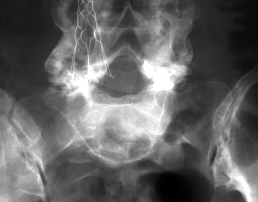
- Morphological Hallmark: The inverted Napoleon hat sign (also referred to as the inverted bicorne hat sign) manifests on frontal anteroposterior (AP) or Ferguson radiographs of the lumbosacral spine and pelvis as a distinctive upside-down cocked hat contour superimposed over the upper sacrum.
- Pathognomonic Pathology: The sign is pathognomonic for severe anterolisthesis (Meyerding Grade III or Grade IV) or complete spondyloptosis (Meyerding Grade V) of the L5 vertebral body over the first sacral segment (S1), typically arising secondary to bilateral isthmic spondylolysis or severe congenital lumbosacral dysplasia.
- Critical Clinical Utility: Identification of the inverted Napoleon hat sign immediately establishes advanced structural lumbosacral deformity, profound sagittal spinopelvic imbalance, and severe traction risk to the exiting L5 spinal nerves, demanding upright whole-spine teleoroentgenograms and prompt specialist surgical evaluation.
2. Diagnostic Sign Summary Matrix
| Primary Modality | Anatomic Location | Primary Pathology | Pathognomonic Morphology | Diagnostic Utility & Sensitivity |
|---|---|---|---|---|
| Plain Radiography (AP / Ferguson) & Coronal CT | Lumbosacral Junction (L5-S1) | High-Grade Spondylolisthesis / Spondyloptosis (Grade III–V) | Upside-down Napoleon hat contour: L5 body forms the downward central crown, transverse processes form the laterally sweeping brim projected over the sacrum | High specificity (>95%) on frontal films; diagnostic hallmark of severe translation and extreme lumbosacral kyphosis |
| Standing Lateral Plain Radiography | Lumbosacral Junction (L5-S1) | Isthmic or Dysplastic Spondylolisthesis | Anterior displacement of L5 body >50% to >100% (spondyloptosis) with sacral dome rounding and elevated slip angle | Gold standard for Meyerding grading (III–V), Dubousset lumbosacral angle, and pelvic tilt measurement |
| Computed Tomography (CT with Multiplanar 2D/3D) | L5 Pars Interarticularis & Sacral Promontory | Bilateral Pars Lysis / Sacral Dome Remodeling | Direct visualization of bony pars gap, trapezoidal wedging of L5 body, and remodeling of the anterosuperior sacral endplate | 100% sensitivity for osseous anatomy and dysplastic facet morphology; critical for screw trajectory planning |
| Magnetic Resonance Imaging (MRI) | L5-S1 Foramina, Lateral Recesses & Cauda Equina | Extraforaminal L5 Root Stretch & S1 Traction | Elongation and drape of L5 spinal nerves over the rounded sacral promontory, pseudo-disc herniation, and central canal crowding | Delineates neural impingement, cauda equina compression, and adjacent-segment disc desiccation at L4-L5 |
3. Clinical Case Imaging & Standardized Figures

4. Pathophysiology & Biomechanical Mechanism
The inverted Napoleon hat sign is a classic projectional radiographic phenomenon that arises directly from the three-dimensional geometric displacement of the fifth lumbar vertebra relative to the sacrum. In physiological alignment, the L5 vertebral body rests squarely atop the S1 superior endplate, with its transverse processes projecting laterally into the iliolumbar spaces. On a standard frontal anteroposterior (AP) radiograph, the L5 body projects as a rectangular contour superior to the pelvic brim, well clear of the sacral wings.
When bilateral structural failure of the pars interarticularis (isthmic spondylolysis) or severe congenital dysplasia of the posterior facets occurs, the neural arch uncouples from the anterior vertebral column. Under sustained physiological gravitational axial loads and the pronounced anterior shear vector inherent to the inclined sacral plateau (typically 40° to 50° in lordotic postures), the L5 body translates anteriorly and pivots caudally into marked flexion. As the anterolisthesis progresses beyond 50% (Meyerding Grade III, IV, or Grade V spondyloptosis), the L5 vertebral body drops anterior and inferior to the sacral promontory, adopting a vertical or steeply kyphotic orientation.
Consequently, an anterior-to-posterior X-ray beam traverses the vertically tilted L5 vertebra in an axial plane, viewing the vertebral body end-on. This geometric orientation projects the dense cortices of the tilted L5 anterior and superior endplates as a smooth, rounded, convex inferior margin, representing the central crown of the inverted cocked hat. Simultaneously, the intact bilateral transverse processes of L5, which maintain continuity with the anterior vertebral cortex, sweep superiorly and laterally toward the iliac crests, forming the curved brim of the upside-down Napoleon hat superimposed directly across the upper sacral segments and sacral ala.
5. Modality-Specific Imaging Findings
Plain Radiography
Plain radiography represents the initial diagnostic cornerstone. On the frontal AP view or 30° to 35° cephalad-angled Ferguson view, the pathognomonic inverted Napoleon hat contour is observed projected over the upper sacrum. The normal L5-S1 disc interspace is obliterated, and the L5 spinous process may appear detached or displaced superiorly due to pars lysis. On the standing lateral lumbar radiograph, the true severity of translation is exposed. The Meyerding grading system classifies the slip based on the percentage of anterior displacement of L5 relative to the anteroposterior diameter of the S1 endplate: Grade III represents 51% to 75% translation, Grade IV represents 76% to 100% translation, and Grade V (spondyloptosis) represents complete translation beyond 100%, where the L5 body sits completely anterior and inferior to the sacral promontory. Associated lateral findings include marked lumbosacral kyphosis (high slip angle), rounding or dome remodeling of the anterior sacrum, and trapezoidal wedging of the L5 vertebral body.
Computed Tomography (CT)
High-resolution multidetector CT with multiplanar coronal, sagittal, and 3D surface reconstructions provides definitive evaluation of the bony architecture. Sagittal CT reformations clearly demonstrate the discontinuous pars interarticularis, distinguishing true isthmic lysis from facet elongation or dysplastic facet hypoplasia. Coronal CT reconstructions replicate the inverted Napoleon hat morphology in thin-slice osseous detail, verifying the precise spatial orientation of the transverse processes relative to the sacral ala. Multiplanar CT is essential for preoperative surgical planning, assessing pedicle dimensions, evaluating sacral dome bone stock, and planning transsacral or S2-alar-iliac (S2AI) screw fixation trajectories.
Magnetic Resonance Imaging (MRI) & Ultrasound
Magnetic resonance imaging is mandatory for appraising neural elements and soft-tissue constraints. Sagittal T1- and T2-weighted sequences visualize the severe downward displacement of the thecal sac and cauda equina across the posterior edge of the sacral dome. The L5 spinal nerves are acutely vulnerable as they exit the foramina, becoming stretched and draped tautly over the rounded sacral promontory in the extraforaminal zone. Axial MRI views evaluate canal compromise and differentiate true compressive disc herniation from pseudo-disc bulging caused by severe translational uncoupling. Although diagnostic ultrasound has no role in adult spondyloptosis, high-resolution sonography can identify pars defects or gross lumbosacral malalignment during infant and pediatric spinal screening.
6. Diagnostic Utility, Sensitivity & Specificity
The inverted Napoleon hat sign demonstrates exceptional diagnostic specificity (>95%) on frontal plain radiographs and pelvic scout films for severe high-grade L5-S1 spondylolisthesis and spondyloptosis. Because the geometric confluence of an axially foreshortened L5 body and upwardly sweeping transverse processes occurs exclusively when L5 undergoes profound anterior translation and caudal tilting over the sacral promontory, false-positive observations in an anatomically normal spine are virtually non-existent.
Diagnostic sensitivity on standard non-targeted abdominal or pelvic radiographs ranges between 75% and 85%, primarily influenced by overlying bowel gas, fecal loading, patient obesity, or suboptimal X-ray beam angulation. In supine non-weight-bearing examinations, partial postural reduction may slightly diminish the prominence of the transverse process brim, although fixed high-grade deformity remains readily identifiable. The sign holds particular clinical utility as a serendipitous alert on non-dedicated abdominal, pelvic, or intravenous urography examinations, signaling massive occult spinal instability to the reviewing clinician.
7. Differential Diagnosis & Radiographic Mimics
| Condition / Sign | Frontal Projection Morphology | Lateral Projection Characteristics | Clinical Presentation & Gold Standard |
|---|---|---|---|
| High-Grade Spondylolisthesis / Spondyloptosis (Classic Sign) | Pathognomonic inverted Napoleon hat contour superimposed over upper sacrum; obliterated L5-S1 disc shadow | Meyerding Grade III–V anterior slip (>50% to >100%); high slip angle; rounded sacral dome; pars defect | Adolescents or adults with crouch gait, hamstring tightness, palpable step-off; confirmed on standing radiographs and CT |
| Low-Grade Spondylolisthesis (Meyerding Grade I–II) | Normal rectangular L5 vertebral body contour; transverse processes project horizontally into iliolumbar space | Anterior displacement <50%; preserved lumbosacral lordosis; absence of significant sacral dome remodeling | Mechanical low back pain; stable spinopelvic balance; confirmed on standing lateral radiography |
| Congenital Lumbosacral Dysplasia / Sacral Agenesis | Asymmetric, hypoplastic, or absent sacral segments; distorted pelvic ring without bicorne hat shadow | Segmentation failure; absence of discrete isthmic pars lysis; variable pelvic dysmorphism | Congenital neurological or pelvic floor deficits; confirmed on whole-spine MRI and 3D pelvic CT |
| Spondylolysis without Spondylolisthesis | Normal vertebral contour; absence of inverted Napoleon hat sign | Intact lumbosacral alignment (Grade 0); pars defect visible on oblique views (“Scottie dog with collar”) | Adolescent hyperextension athlete; localized extension back pain; confirmed on thin-slice CT or SPECT/MRI |
| Destructive Lumbosacral Neoplasm / Infection | Ragged osteolytic bone destruction of L5 or S1; soft-tissue mass obscuring pelvic margins | Pathological collapse or anterior subluxation with cortical erosion and bone marrow replacement | Severe constant pain, night pain, constitutional symptoms, elevated ESR/CRP; confirmed on contrast MRI and biopsy |
8. Clinical Significance & Management Implications
The presence of an inverted Napoleon hat sign signifies end-stage structural incompetence of the lumbosacral junction, necessitating thorough clinical and spinopelvic radiographic assessment:
- Physical Examination Hallmarks: Patients with high-grade slips and spondyloptosis typically present with a characteristic physical appearance: foreshortening of the trunk with a transverse abdominal crease, flattened buttocks due to vertical pelvic orientation, a prominent palpable step-off above the sacrum, and the classic Phalen-Dickson gait (crouch gait with flexed hips and knees secondary to severe hamstring spasticity).
- Spinopelvic Sagittal Balance Assessment: According to the Spinal Deformity Study Group (SDSG) classification, high-grade slips (Types 4, 5, and 6) are categorized by pelvic tilt and global sagittal vertical axis (SVA). Patients with a balanced pelvis (low pelvic tilt, Type 4) experience less compensatory fatigue than those with a retroverted, unbalanced pelvis (high pelvic tilt, Type 5) or uncompensated global sagittal imbalance (Type 6), directly governing the magnitude of realignment required.
- Surgical Indications & Decompression: Conservative therapy is rarely definitive in progressive high-grade slips. Surgical intervention is indicated for progressive slip, intractable mechanical back pain, radiculopathy, or cauda equina compromise. Wide laminectomy (Gill procedure) with extensive unroofing of the exiting L5 roots is standard.
- Reduction versus In Situ Fusion Dilemma: In situ fusion carries a low risk of neurological injury but leaves the severe lumbosacral kyphosis uncorrected, predisposing patients to progressive deformity, high pseudarthrosis rates, and flatback syndrome. Conversely, complete anatomical reduction subjects the stretched L5 nerve root to acute traction neuropathy. Contemporary surgical consensus favors partial reduction focused on correcting the slip angle (lumbosacral kyphosis) rather than translational displacement, often paired with posterior column shortening (L5 partial spondylectomy or sacral dome resection) and circumferential rigid multi-rod fixation with pelvic S2AI anchors.
9. Master Class Pro-Tip
Master Class Pro-Tip: When evaluating a patient with the inverted Napoleon hat sign on frontal imaging, never attempt full translational reduction of the L5 vertebra onto the sacral plateau without first addressing the slip angle and lumbosacral kyphosis. The exiting L5 spinal nerve is acutely tethered over the rounded, remodeled sacral dome in the extraforaminal gutter. Vigorous posterior translational reduction subjects this tethered L5 nerve root to severe traction ischemia, precipitating postoperative foot drop. Instead, prioritize correcting the lumbosacral kyphosis to restore global spinopelvic sagittal balance, and perform intentional posterior column shortening or partial sacral dome resection before translational docking.
Master Class Radiological Pearl: Sagittal Realignment and Nerve Root Protection in Spondyloptosis
10. Evidence-Based Synthesis
| Author & Year | Study Design | Sample Size | Key Findings & Diagnostic Accuracy | Clinical Implication | Level of Evidence | DOI |
|---|---|---|---|---|---|---|
| Garland LH et al. (1946) | Landmark Radiographic Investigation | N = 100 spine radiographs | First documented radiological description of the inverted Napoleon hat contour produced on frontal radiographs by the severe anterior and caudal displacement of L5 superimposed over the upper sacrum. | Established the pathognomonic diagnostic sign for severe anterolisthesis and spondyloptosis of L5 on S1. | Level V | PubMed |
| Talangbayan LE (2007) | Classic Radiological Sign Review | N = 1 (Review of sign mechanics) | Detailed the exact radiographic geometry: anterolisthesis and vertical tilting of L5 causes the anterior body margin to form the crown and the transverse processes to form the brim of an inverted bicorne hat on AP projection. | Confirmed the pathognomonic nature of the sign for severe Grade III to V spondylolisthesis on standard frontal abdominal or pelvic radiographs. | Level V | DOI |
| Pendergrast TE et al. (2019) | Abdominal & Pelvic Imaging Review | N = 1 (Case & Diagnostic Review) | Demonstrated that the inverted Napoleon hat sign can be rapidly recognized on non-dedicated pelvic radiographs and abdominal scout views, alerting clinicians to severe occult lumbosacral instability. | Enables non-spine specialists to promptly identify severe lumbosacral malalignment on routine abdominal examinations. | Level V | DOI |
| Lak AM et al. (2020) | Systematic Review & Meta-Analysis | N = 312 patients across 14 studies | Systematic comparison showed reduction of high-grade slips achieved superior restoration of slip angle and spinopelvic balance with higher fusion rates, balanced against a transiently increased risk of L5 radiculopathy. | Guides decision-making between partial reduction to correct sagittal balance versus in situ fusion in adult high-grade spondylolisthesis. | Level I | DOI |
| Mac-Thiong JM et al. (2012) | Multicenter Reliability Study | N = 126 patients | Validated the SDSG classification for high-grade spondylolisthesis (slip > 50%), stratifying patients into balanced vs. unbalanced pelvis and spine to direct surgical alignment strategies. | Establishes standard nomenclature and global sagittal balance criteria for cases presenting with inverted Napoleon hat deformity. | Level II | DOI |
| Labelle H et al. (2011) | Biomechanical Classification & Review | N = 214 patients | Demonstrated that high-grade slips are characterized by high pelvic incidence, retroverted pelvis, and focal lumbosacral kyphosis, which trigger compensatory hip flexion and knee flexion. | Highlights that correcting the lumbosacral kyphosis (slip angle) is essential for restoring upright sagittal posture. | Level IV | DOI |
| Camino Willhuber G et al. (2020) | Expert Classification Review | Review | Summarized the SDSG system dividing high-grade slips (Types 4 to 6) based on pelvic tilt and sagittal vertical axis, demonstrating high diagnostic and surgical concordance. | Provides practical guidance for choosing between in situ fusion, reduction, or postural realignment based on spinopelvic parameters. | Level V | DOI |
| Obeid I et al. (2015) | Retrospective Comparative Surgical Series | N = 23 patients | Posterior column shortening via partial L5 vertebrectomy enabled safe anatomical reduction, reducing slip angle from 38° to 4° without permanent L5 nerve root injury. | Demonstrates that intentional posterior column shortening protects the L5 nerve root during reduction of severe high-grade spondylolisthesis and spondyloptosis. | Level IV | DOI |
| Viroli G et al. (2024) | Retrospective Comparative Cohort Study | N = 45 patients | Surgical reduction significantly improved pelvic tilt, sacral slope, and lumbar lordosis, achieving normal spinopelvic balance in 82% of patients with high-grade slips. | Confirmed durable correction of compensatory posture following anatomical reduction and circumferential fusion in high-grade spondylolisthesis. | Level III | DOI |
11. References
- Garland LH, Thomas SF. Spondylolisthesis; criteria for more accurate diagnosis of true anterior slip of the involved vertebral segment. Am J Roentgenol Radium Ther. 1946;55(3):275-291. PubMed [Level V: Landmark Radiographic Investigation]
- Talangbayan LE. The Inverted Napoleon’s Hat Sign. Radiology. 2007;243(2):603-604. DOI [Level V: Classic Radiological Sign Review]
- Pendergrast TE, Dyer RB. The “inverted Napoleon’s hat” sign. Abdom Radiol (NY). 2019;44(8):2936-2937. DOI [Level V: Abdominal & Pelvic Imaging Review]
- Lak AM, Abunimer AM, Devi S, et al. Reduction versus In Situ Fusion for Adult High-Grade Spondylolisthesis: A Systematic Review and Meta-Analysis. World Neurosurg. 2020;138:e791-e799. DOI [Level I: Systematic Review & Meta-Analysis]
- Mac-Thiong JM, Duong L, Parent S, et al. Reliability of the Spinal Deformity Study Group Classification of Lumbosacral Spondylolisthesis. Spine. 2012;37(2):E95-E102. DOI [Level II: Multicenter Reliability Study]
- Labelle H, Mac-Thiong JM, Roussouly P. Spino-pelvic sagittal balance of spondylolisthesis: a review and classification. Eur Spine J. 2011;20(Suppl 5):641-646. DOI [Level IV: Biomechanical Classification & Review]
- Camino Willhuber G, Kido G. Classifications in Brief: The Spinal Deformity Study Group Classification of Lumbosacral Spondylolisthesis. Clin Orthop Relat Res. 2020;478(4):898-904. DOI [Level V: Expert Classification Review]
- Obeid I, Laouissat F, Bourghli A, et al. One-stage posterior spinal shortening by L5 partial spondylectomy for spondyloptosis or L5-S1 high-grade spondylolisthesis management. Eur Spine J. 2015;24(10):2220-2228. DOI [Level IV: Retrospective Comparative Surgical Series]
- Viroli G, Ruffilli A, Ialuna M, et al. Restoration of Spinopelvic Alignment After Reduction of High-grade Spondylolisthesis. Clin Spine Surg. 2024;37(4):171-177. DOI [Level III: Retrospective Comparative Cohort Study]
Last Updated on September 28, 2026
🔗 Knowledge Graph & Clinical Correlates
OrthopaedicsOne Living Semantic Network🛠️ Operative Techniques
🩺 Clinical Conditions & Indications
- Torts 1. High-Yield Summary Medical tort liability in orthopaedic surgery arises when clinical care departs from accepted standards of...
- Timing of secondary sexual characteristics 1. High-Yield Overview & Clinical Purpose Biological Staging and Pubertal Timing: Secondary sexual characteristics, categorized via the Marshall...
- Thoracolumbar trauma Introduction Trauma to the lumbar or thoracic spine remains a clinical challenge from a diagnostic and management perspective....
- Thoracolumbar sprains 1. High-Yield Summary Definition & Core Distinction: Thoracolumbar sprains involve mechanical injury, micro-tearing, or partial disruption of the...
🔬 Anatomy, Pathology & Diagnostics
- Vertebroplasty and Kyphoplasty for Osteolytic Vertebral Collapse Lumbar Spine
- Vertebral endplate and marrow changes – Modic classification Lumbar Spine
- Vertebra prominens Surface Anatomy Lumbar Spine
- Tumbling Bullet Sign Lumbar Spine